File:Fetal blood flow 01.jpg: Difference between revisions
| Line 1: | Line 1: | ||
==Fetal Blood Flow== | ==Fetal Blood Flow== | ||
Mean flows in the major vessels of the human fetal circulation by phase contrast MRI. Mean flows in ml/kg/min | Mean flows in the major vessels of the human fetal circulation by phase contrast MRI. | ||
* '''left''' - Mean flows in ml/kg/min | |||
* '''right''' - Proportions of the combined ventricular output in the major vessels of the human fetal circulation by phase contrast MRI. | |||
Ascending aorta (AAo), | |||
main pulmonary artery (MPA), ductus arteriosus (DA), pulmonary blood flow (PBF), descending aorta (DAo), umbilical artery (UA), umbilical vein (UV), inferior vena cava (IVC), superior vena cava (SVC), right atrium (RA), foramen ovale (FO), left atrium (LA), right ventricle (RV), left ventricle (LV). | |||
===Reference=== | ===Reference=== | ||
| Line 16: | Line 21: | ||
Figure 2. | |||
Journal of Cardiovascular Magnetic Resonance 2012, 14:79 doi:10.1186/1532-429X-14-79 | |||
[[Category:Human]] [[Category:Cardiovascular]] [[Category:MRI]] | [[Category:Human]] [[Category:Cardiovascular]] [[Category:MRI]] | ||
Revision as of 16:26, 3 January 2013
Fetal Blood Flow
Mean flows in the major vessels of the human fetal circulation by phase contrast MRI.
- left - Mean flows in ml/kg/min
- right - Proportions of the combined ventricular output in the major vessels of the human fetal circulation by phase contrast MRI.
Ascending aorta (AAo),
main pulmonary artery (MPA), ductus arteriosus (DA), pulmonary blood flow (PBF), descending aorta (DAo), umbilical artery (UA), umbilical vein (UV), inferior vena cava (IVC), superior vena cava (SVC), right atrium (RA), foramen ovale (FO), left atrium (LA), right ventricle (RV), left ventricle (LV).
Reference
<pubmed>23181717</pubmed>| J Cardiovasc Magn Reson.
Copyright=
© 2012 Seed et al.; licensee BioMed Central Ltd. This is an Open Access article distributed under the terms of the Creative Commons Attribution License ( http://creativecommons.org/licenses/by/2.0), which permits unrestricted use, distribution, and reproduction in any medium, provided the original work is properly cited.
Figure 2.
Journal of Cardiovascular Magnetic Resonance 2012, 14:79 doi:10.1186/1532-429X-14-79
File history
Click on a date/time to view the file as it appeared at that time.
| Date/Time | Thumbnail | Dimensions | User | Comment | |
|---|---|---|---|---|---|
| current | 16:22, 3 January 2013 | 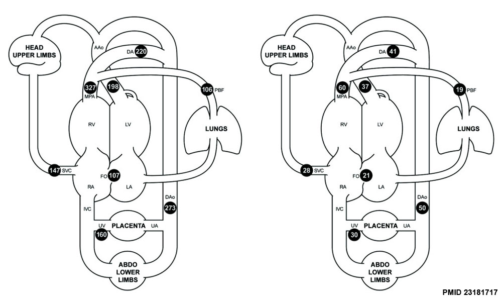 | 1,000 × 599 (75 KB) | Z8600021 (talk | contribs) | ==Fetal Blood Flow== Mean flows in the major vessels of the human fetal circulation by phase contrast MRI. Mean flows in ml/kg/min (left) and proportions of the combined ventricular output (right) in the major vessels of the human fetal circulation by ph |
You cannot overwrite this file.
File usage
The following 3 pages use this file: