File:Fetal Colonic Inury Model Diagram.png: Difference between revisions
(Diagram of Fetal Injury Colonic Model as seen in study: PMID: 24139758) |
No edit summary |
||
| Line 1: | Line 1: | ||
==Fetal Intestine Transplantat Injury Model== | |||
This is a hand drawn digram of the process described in the study by R. Fordham et. al <ref name="PMID3858813"><pubmed>3858813</pubmed></ref>. The left hand side of the diagram shows the immature cells of the human body (induced pluripotent cells shown in red and the immature cells of the intestine shown down the bottom in the red and blue) that can form spondin independent Fetal Enterospheres (FEnS). This is important as in this study using models of both fetal mouse and humans it was found that FEnS contribute to the regeneration of colonic epithelium by the formation of drypt-like epithelial structures that expressed region-specific differentiation markers<ref name="PMID3858813"><pubmed>3858813</pubmed></ref>. The right hand side of the diagram shows the mature intestine cells. From differing techniques it was found that the FEnS were able to form regeneration of an intestinal injury while in vitro maturation of the immature intestinal cells merely formed the spondin dependant organoids. | |||
==References== | |||
<references/> | |||
{{Template:Student Image}} | |||
Revision as of 17:45, 23 October 2014
Fetal Intestine Transplantat Injury Model
This is a hand drawn digram of the process described in the study by R. Fordham et. al [1]. The left hand side of the diagram shows the immature cells of the human body (induced pluripotent cells shown in red and the immature cells of the intestine shown down the bottom in the red and blue) that can form spondin independent Fetal Enterospheres (FEnS). This is important as in this study using models of both fetal mouse and humans it was found that FEnS contribute to the regeneration of colonic epithelium by the formation of drypt-like epithelial structures that expressed region-specific differentiation markers[1]. The right hand side of the diagram shows the mature intestine cells. From differing techniques it was found that the FEnS were able to form regeneration of an intestinal injury while in vitro maturation of the immature intestinal cells merely formed the spondin dependant organoids.
References
- Note - This image was originally uploaded as part of an undergraduate science student project and may contain inaccuracies in either description or acknowledgements. Students have been advised in writing concerning the reuse of content and may accidentally have misunderstood the original terms of use. If image reuse on this non-commercial educational site infringes your existing copyright, please contact the site editor for immediate removal.
File history
Click on a date/time to view the file as it appeared at that time.
| Date/Time | Thumbnail | Dimensions | User | Comment | |
|---|---|---|---|---|---|
| current | 17:08, 23 October 2014 | 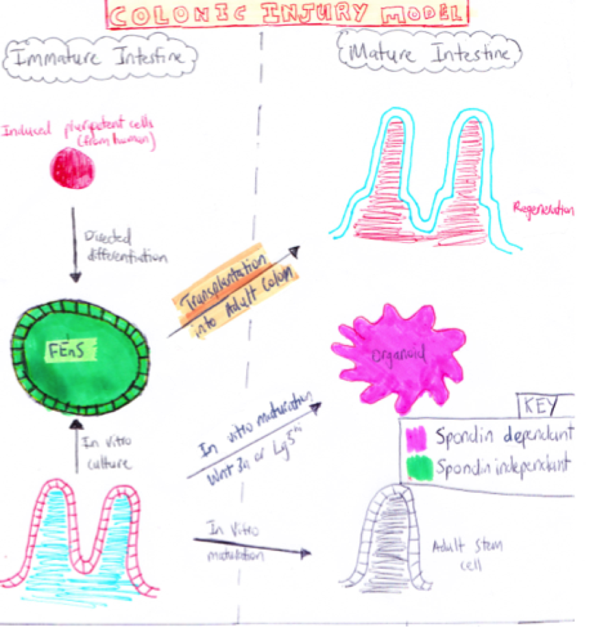 | 865 × 916 (650 KB) | Z3415141 (talk | contribs) | Diagram of Fetal Injury Colonic Model as seen in study: PMID: 24139758 |
You cannot overwrite this file.
File usage
The following 2 pages use this file: