File:Fetal Circulation Pathway.jpg

Original file (1,371 × 1,069 pixels, file size: 112 KB, MIME type: image/jpeg)
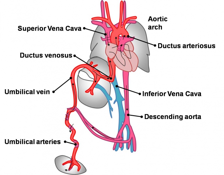
Fetal Circulation
Fetal circulation consequently differs from the adult one predominantly due to the presence of 3 major vascular shunts:
- Ductus venosus - between the umbilical vein and IVC
- Foramen ovale - between the right and left atrium
- Ductus arteriosus - between the pulmonary artery and descending aorta
The main function of these shunts is to redirect oxygenated blood away from the lungs, liver and kidney (whose functions are performed by the placenta).
Oxygenated blood is carried from the placenta to the foetus in the umbilical vein, most of which then passes through the ductus venosus to the IVC while some blood supplies the liver via the portal vein. Blood from the liver drains into the IVC through the hepatic veins. The blood in the IVC is a mixture of oxygenated blood from the umbilical vein and desaturated blood from the lower limbs and abdominal organs (e.g. the liver). This blood enters the right atrium where most of it is directed to the left atrium through the foramen ovale and from here to the left ventricle and aorta. The remainder of the blood in the right atrium passes with blood from the SVC (from the head and upper limbs) to the right ventricle and pulmonary artery where most of it passes to the aorta via the ductus arteriosus. The blood passes from the aorta to the hypogastric arteries, umbilical arteries and then back to the placenta.
- Links:
File history
Click on a date/time to view the file as it appeared at that time.
| Date/Time | Thumbnail | Dimensions | User | Comment | |
|---|---|---|---|---|---|
| current | 10:29, 14 March 2010 | | 1,371 × 1,069 (112 KB) | Z3212774 (talk | contribs) | category:Heart ILP |
You cannot overwrite this file.
File usage
The following 3 pages use this file: