File:Fawcett 1910 fig12.jpg: Difference between revisions
({{Fawcett1910_sphenoid_figures}}) |
mNo edit summary |
||
| Line 1: | Line 1: | ||
==Fig. 12 Coronal section of the head of a 37 mm embryo Harvard Collection== | |||
[[Harvard Collection]] It shows us that the external pterygoidplate (E.P.) is composed of connective tissue. E.P.M.is the external pterygoid muscle; I.P.P. is the bony (ectochondral) internal pterygoid plate; T.P. the tensor palati; M. mandible; M.C. Meckel's cartilage; M.G. Meckel's ganglion; N.S. nasal septum; N. nasal cavity; 0. the mouth; I.P. internal pterygoid muscle; P. the ununited palate. | |||
{{Fawcett1910_sphenoid_figures}} | {{Fawcett1910_sphenoid_figures}} | ||
[[Category:Harvard Collection]] | |||
Latest revision as of 08:57, 29 December 2014
Fig. 12 Coronal section of the head of a 37 mm embryo Harvard Collection
Harvard Collection It shows us that the external pterygoidplate (E.P.) is composed of connective tissue. E.P.M.is the external pterygoid muscle; I.P.P. is the bony (ectochondral) internal pterygoid plate; T.P. the tensor palati; M. mandible; M.C. Meckel's cartilage; M.G. Meckel's ganglion; N.S. nasal septum; N. nasal cavity; 0. the mouth; I.P. internal pterygoid muscle; P. the ununited palate.
| Historic Disclaimer - information about historic embryology pages |
|---|
 |
|
|
Reference
Fawcett E. Notes on the development of the human sphenoid. (1910) J Anat. Physiol. 44(3): 207-22. PMID 17232842
Cite this page: Hill, M.A. (2024, April 16) Embryology Fawcett 1910 fig12.jpg. Retrieved from https://embryology.med.unsw.edu.au/embryology/index.php/File:Fawcett_1910_fig12.jpg
- © Dr Mark Hill 2024, UNSW Embryology ISBN: 978 0 7334 2609 4 - UNSW CRICOS Provider Code No. 00098G
File history
Click on a date/time to view the file as it appeared at that time.
| Date/Time | Thumbnail | Dimensions | User | Comment | |
|---|---|---|---|---|---|
| current | 08:33, 29 December 2014 | 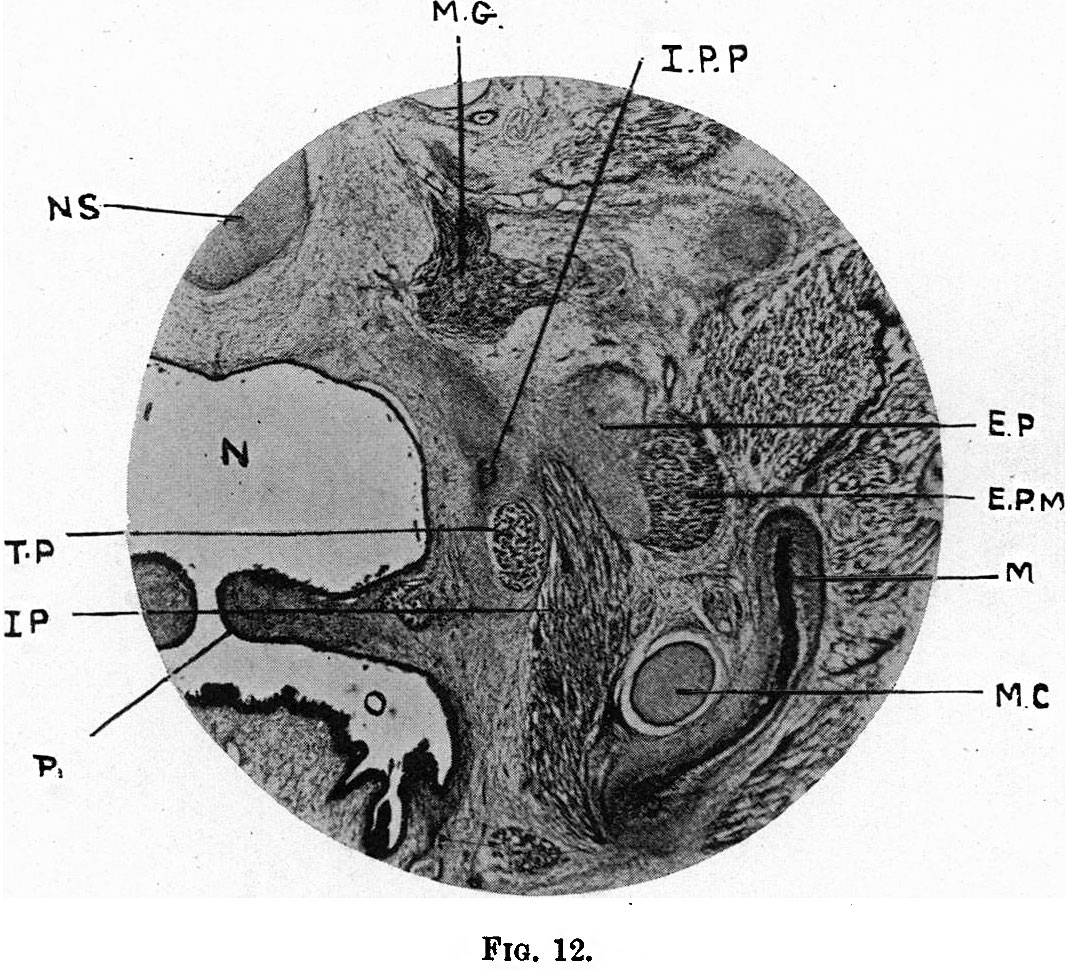 | 1,068 × 970 (282 KB) | Z8600021 (talk | contribs) | {{Fawcett1910_sphenoid_figures}} |
You cannot overwrite this file.
File usage
The following page uses this file: