File:Fawcett 1910 fig11.jpg

Original file (768 × 719 pixels, file size: 124 KB, MIME type: image/jpeg)
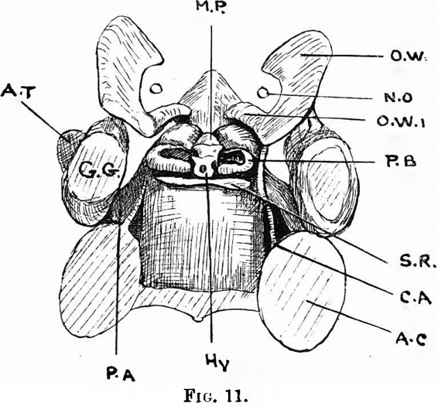
Fig.11. Coronal section of a 19 mm Embryo
View of the same model from above.
O.W. the orbital wing, with O.W.1, its posterior limb, lying behind the optic nerve and running forwards by the side of the prepituitary segment of the corpus sphenoidale. This is the part marked O.W.1 in the photographs 8 and 9. We see here, too, the middle piece (M.P.), which is thus evident both above and below,butis not an independent formation. P.B. is the pituitary body; Hy. the hypophysis; S.R. is the saddle ridge "Sattellehne" ordorsumn selav and it is important to note, first, that it is a rod of cartilage transversely, pltaed, and, second, that it is quite independent of the rest at this stage; A.T. isthe ala teInporalis seen under cover of G.G., the Gassernia ganglion; C.A. is the carotid artery; A.C. the auditory capsule; whilst P.A. isthe processus alaris.
| Historic Disclaimer - information about historic embryology pages |
|---|
 |
|
|
Reference
Fawcett E. Notes on the development of the human sphenoid. (1910) J Anat. Physiol. 44(3): 207-22. PMID 17232842
Cite this page: Hill, M.A. (2024, April 23) Embryology Fawcett 1910 fig11.jpg. Retrieved from https://embryology.med.unsw.edu.au/embryology/index.php/File:Fawcett_1910_fig11.jpg
- © Dr Mark Hill 2024, UNSW Embryology ISBN: 978 0 7334 2609 4 - UNSW CRICOS Provider Code No. 00098G
File history
Click on a date/time to view the file as it appeared at that time.
| Date/Time | Thumbnail | Dimensions | User | Comment | |
|---|---|---|---|---|---|
| current | 08:33, 29 December 2014 | | 768 × 719 (124 KB) | Z8600021 (talk | contribs) | {{Fawcett1910_sphenoid_figures}} |
You cannot overwrite this file.
File usage
The following page uses this file: