The printable version is no longer supported and may have rendering errors. Please update your browser bookmarks and please use the default browser print function instead.
| Historic Disclaimer - information about historic embryology pages
|
| Pages where the terms "Historic" (textbooks, papers, people, recommendations) appear on this site, and sections within pages where this disclaimer appears, indicate that the content and scientific understanding are specific to the time of publication. This means that while some scientific descriptions are still accurate, the terminology and interpretation of the developmental mechanisms reflect the understanding at the time of original publication and those of the preceding periods, these terms, interpretations and recommendations may not reflect our current scientific understanding. (More? Embryology History | Historic Embryology Papers)
|
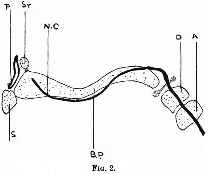
- A. Anterior limb of ala orbitalis forming anterior wall of optic foramen.
- P. Posterior limb of ala orbitalis forming the posterior and inner walls of the optic foramen.
- S.E.C. The spheno-ethmoidal cartilage.
- A.O. Outer end of the spheno-ethmoidal cartilage extending into the temporal fosa.
- I.O.N. Infraorbital nerve.
- M.G. Meckel's ganglion.
|
- A.T. Ala temporalis.
- M. Maxilla.
- N.C. Nasal capsule.
- C. Connective tissue in which nasal bone ossifies together with part of the nasal process of the maxilla.
- L.D. Lacrymal duct.
- L.N.P. Lateral nasal cartilage (MAlihalkowics). Ma. Malarbone
- P.P. Parietal plate.
- F. Frontal bone.
|
- Sphenoid Links: Fig. 1. Fig. 2.
Reference
Fawcett E. Notes on the development of the human sphenoid. (1910) J Anat. Physiol. 44(3): 207-22. PMID 17232842
Cite this page: Hill, M.A. (2024, April 16) Embryology Fawcett 1910 fig02.jpg. Retrieved from https://embryology.med.unsw.edu.au/embryology/index.php/File:Fawcett_1910_fig02.jpg
- What Links Here?
- © Dr Mark Hill 2024, UNSW Embryology ISBN: 978 0 7334 2609 4 - UNSW CRICOS Provider Code No. 00098G
File history
Click on a date/time to view the file as it appeared at that time.
| Date/Time | Thumbnail | Dimensions | User | Comment |
|---|
| current | 08:30, 29 December 2014 |  | 824 × 690 (56 KB) | Z8600021 (talk | contribs) | {{Fawcett1910_sphenoid_figures}} |
You cannot overwrite this file.
File usage
The following page uses this file:
This file contains additional information, probably added from the digital camera or scanner used to create or digitise it.
If the file has been modified from its original state, some details may not fully reflect the modified file.

