File:Fawcett1910 fig03.jpg: Difference between revisions
From Embryology
mNo edit summary |
mNo edit summary |
||
| Line 2: | Line 2: | ||
The cartilage is everywhere "stippled." | The cartilage is everywhere "stippled." | ||
* '''A.C.''' - auditory capsule | |||
* '''A.T.''' - ala temporalis or pars zygomatica (the third division of the 5th nerve is seen crossing its outerside) | |||
* '''C.''' - cricoid cartilage | |||
* '''C.Ty.''' - chorda tympani nerve | |||
* '''C2 - neural arch of second cervical vertebra | |||
* '''C3 - neural arch of third cervical vertebra | |||
* '''C.B.''' - connective tissue bar which forms part of outer wall of orbit which will later ossify to complete the malar bone and external angular process of the frontal bone | |||
* '''E.P.''' - ethmoidal plate | |||
* '''F.''' - frontal bone | |||
* '''I.C.''' - internal carotid artery | |||
* '''H.''' - cerato- - epi- - stylo- and tympano-hyal | |||
* '''G.O.N.''' - great occipital nerve passing backwards from second cervical nerve | |||
* '''L.D.''' - lacrymal duct;L.N.P. lateral nasal process | |||
* '''Ma.''' - malar bone (behind and deep to it the palate bone is seen) | |||
* '''Mn.''' - mandible with Meckel's cartilage (Mt.) running backwards from it and forming malleus cartilage | |||
* '''Mx.''' - maxilla - showing the infraorbital groove through which the infraorbital nerve is running | |||
* '''N.C.''' - nasal capsule | |||
* '''O.P.''' - ala orbitalis or lesser wing pointing backwards towards P.P.''' - the parietal plate | |||
* '''P.P.''' - parietal plate | |||
* '''S.O.M.''' - spino-occipital membrane | |||
* '''S.Z.''' - squamoso-zygomatic | |||
* '''T.S.''' - tectum synoticum | |||
* '''T.F.''' - temporal fossa limited behind and above by a groove | |||
* '''V.''' - vacuity caused by large vein or extravasation | |||
* '''V.S.''' - vagus and sympathetic ganglion. | |||
{{Fawcett1910 figures}} | {{Fawcett1910 figures}} | ||
Revision as of 06:34, 27 December 2014
Fig. 3. Profile view of neuro-cranium, visceral cranium, and upper part of neck skeleton
The cartilage is everywhere "stippled."
- A.C. - auditory capsule
- A.T. - ala temporalis or pars zygomatica (the third division of the 5th nerve is seen crossing its outerside)
- C. - cricoid cartilage
- C.Ty. - chorda tympani nerve
- C2 - neural arch of second cervical vertebra
- C3 - neural arch of third cervical vertebra
- C.B. - connective tissue bar which forms part of outer wall of orbit which will later ossify to complete the malar bone and external angular process of the frontal bone
- E.P. - ethmoidal plate
- F. - frontal bone
- I.C. - internal carotid artery
- H. - cerato- - epi- - stylo- and tympano-hyal
- G.O.N. - great occipital nerve passing backwards from second cervical nerve
- L.D. - lacrymal duct;L.N.P. lateral nasal process
- Ma. - malar bone (behind and deep to it the palate bone is seen)
- Mn. - mandible with Meckel's cartilage (Mt.) running backwards from it and forming malleus cartilage
- Mx. - maxilla - showing the infraorbital groove through which the infraorbital nerve is running
- N.C. - nasal capsule
- O.P. - ala orbitalis or lesser wing pointing backwards towards P.P. - the parietal plate
- P.P. - parietal plate
- S.O.M. - spino-occipital membrane
- S.Z. - squamoso-zygomatic
- T.S. - tectum synoticum
- T.F. - temporal fossa limited behind and above by a groove
- V. - vacuity caused by large vein or extravasation
- V.S. - vagus and sympathetic ganglion.
Links: Fig 1 | Fig 2 | Fig 3 | Fig 4 | Skull Development | Carnegie stage 23
| Historic Disclaimer - information about historic embryology pages |
|---|
 |
- Edward Fawcett Links: 1906 Palate | 1910 Head | 1910 Sphenoid | 1911 Maxilla, vomer, and paraseptal cartilages | 1913 Clavicle | 1930 Mandible | Fawcett image | Edward Fawcett
Reference
Fawcett E. Description of a reconstruction of the head of a thirty-millimetre embryo. (1910) J Anat. Physiol. 44(4): 303-11.
Cite this page: Hill, M.A. (2024, April 24) Embryology Fawcett1910 fig03.jpg. Retrieved from https://embryology.med.unsw.edu.au/embryology/index.php/File:Fawcett1910_fig03.jpg
- © Dr Mark Hill 2024, UNSW Embryology ISBN: 978 0 7334 2609 4 - UNSW CRICOS Provider Code No. 00098G
File history
Click on a date/time to view the file as it appeared at that time.
| Date/Time | Thumbnail | Dimensions | User | Comment | |
|---|---|---|---|---|---|
| current | 06:12, 27 December 2014 |  | 905 × 978 (160 KB) | Z8600021 (talk | contribs) | |
| 06:11, 27 December 2014 | 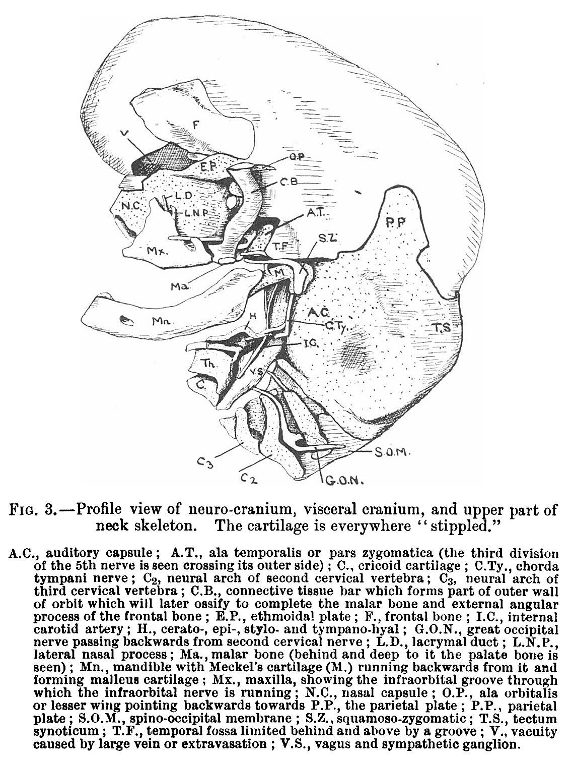 | 1,136 × 1,507 (416 KB) | Z8600021 (talk | contribs) |
You cannot overwrite this file.
File usage
The following 2 pages use this file: