File:Fawcett1910 fig01.jpg: Difference between revisions
From Embryology
No edit summary |
mNo edit summary |
||
| (4 intermediate revisions by the same user not shown) | |||
| Line 1: | Line 1: | ||
==Fig. 1. Part of the reconstructed head of the Bryce 30 mm embryo as seen from the front and left side== | |||
* '''A.''' - ant. limb of ala orbitalis | |||
* '''A.O.''' - outer end of ala orbitalis of sphenoid | |||
* '''A.T.''' - ala tenmporalis''' - perforated by superior maxillary nerve | |||
* '''C.''' - connective tissue bar over nasal capsule in which nasal bones and internal angular processes of frontal bones will develop | |||
* '''F.''' - frontal bonie | |||
* '''L.D.''' - lacrymal duct | |||
* '''L.N.P.''' - lateral nasal process | |||
* '''I.O.N.''' - infraorbital nerve | |||
* '''M.''' - maxilla | |||
* '''Ma.''' - malar bone at lower end of vertical bar of connective tissue which is perforated by temporo-malar nerve | |||
* '''M.G.''' - Meckel's ganglion | |||
* '''O.P.''' - part of outer wall of orbit formed by connective tissue and which later ossifies to form the orbital plate of the great wing of the sphenoid | |||
* '''P.''' - posterior limb of ala orbitalis (lesser wing of sphenoid) between A and P notice optic nerve. | |||
* '''P.P.''' - parietal plate | |||
* '''S.E.C.''' - ethmoidal or spheno-ethmoidal plate. | |||
<br> | |||
{{Carnegie stage 23 links}} | |||
<br> | |||
{{Fawcett1910 figures}} | |||
Latest revision as of 13:23, 22 May 2017
Fig. 1. Part of the reconstructed head of the Bryce 30 mm embryo as seen from the front and left side
- A. - ant. limb of ala orbitalis
- A.O. - outer end of ala orbitalis of sphenoid
- A.T. - ala tenmporalis - perforated by superior maxillary nerve
- C. - connective tissue bar over nasal capsule in which nasal bones and internal angular processes of frontal bones will develop
- F. - frontal bonie
- L.D. - lacrymal duct
- L.N.P. - lateral nasal process
- I.O.N. - infraorbital nerve
- M. - maxilla
- Ma. - malar bone at lower end of vertical bar of connective tissue which is perforated by temporo-malar nerve
- M.G. - Meckel's ganglion
- O.P. - part of outer wall of orbit formed by connective tissue and which later ossifies to form the orbital plate of the great wing of the sphenoid
- P. - posterior limb of ala orbitalis (lesser wing of sphenoid) between A and P notice optic nerve.
- P.P. - parietal plate
- S.E.C. - ethmoidal or spheno-ethmoidal plate.
Links: Fig 1 | Fig 2 | Fig 3 | Fig 4 | Skull Development | Carnegie stage 23
| Historic Disclaimer - information about historic embryology pages |
|---|
 |
- Edward Fawcett Links: 1906 Palate | 1910 Head | 1910 Sphenoid | 1911 Maxilla, vomer, and paraseptal cartilages | 1913 Clavicle | 1930 Mandible | Fawcett image | Edward Fawcett
Reference
Fawcett E. Description of a reconstruction of the head of a thirty-millimetre embryo. (1910) J Anat. Physiol. 44(4): 303-11.
Cite this page: Hill, M.A. (2024, April 16) Embryology Fawcett1910 fig01.jpg. Retrieved from https://embryology.med.unsw.edu.au/embryology/index.php/File:Fawcett1910_fig01.jpg
- © Dr Mark Hill 2024, UNSW Embryology ISBN: 978 0 7334 2609 4 - UNSW CRICOS Provider Code No. 00098G
File history
Click on a date/time to view the file as it appeared at that time.
| Date/Time | Thumbnail | Dimensions | User | Comment | |
|---|---|---|---|---|---|
| current | 06:13, 27 December 2014 | 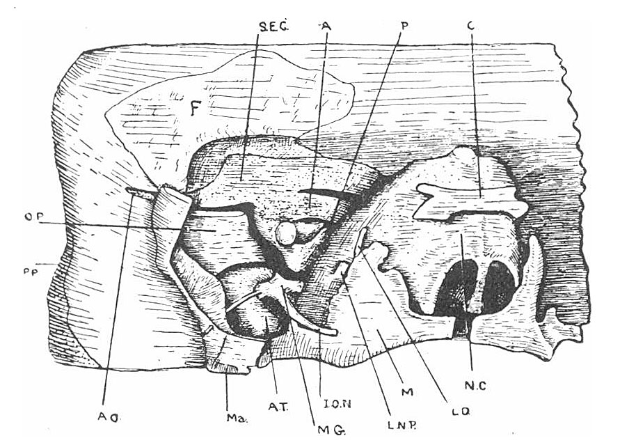 | 891 × 639 (148 KB) | Z8600021 (talk | contribs) | |
| 06:10, 27 December 2014 | 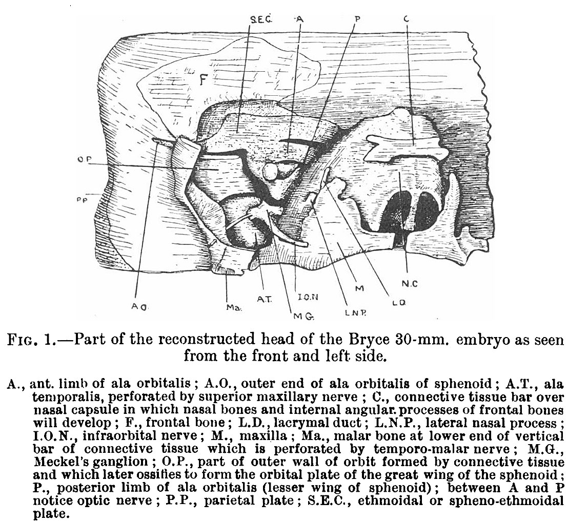 | 1,106 × 1,021 (310 KB) | Z8600021 (talk | contribs) |
You cannot overwrite this file.
File usage
The following page uses this file: