File:Faulconer1951 fig05.jpg
Original file (1,014 × 847 pixels, file size: 233 KB, MIME type: image/jpeg)
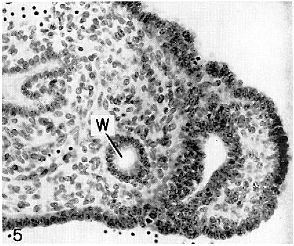
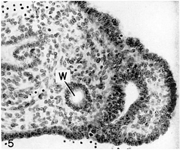
Fig. 5.
Fig. 5. Large Müllerian duct formed by the invagin:1 tion of the ventral surface of the right Wolffian body in the 14.2 mm embryo. Note the smaller Wolffian duct (IV) medial to the Müllerian duct. Embryo no. 6520. X300.
| Historic Disclaimer - information about historic embryology pages |
|---|
 |
Links: fig 1 | plate 1 | fig 2 | fig 3 | fig 4 | fig 5 | fig 6 | fig 7 | Uterus Development | Genital Development
Reference
Falconer RI. Observations on the origin of the Mullerian groove in human embryos. (1951) Carnegie Instn. Wash. Publ. 611, Contrib. Embryol., .
Cite this page: Hill, M.A. (2024, April 18) Embryology Faulconer1951 fig05.jpg. Retrieved from https://embryology.med.unsw.edu.au/embryology/index.php/File:Faulconer1951_fig05.jpg
- © Dr Mark Hill 2024, UNSW Embryology ISBN: 978 0 7334 2609 4 - UNSW CRICOS Provider Code No. 00098G
File history
Click on a date/time to view the file as it appeared at that time.
| Date/Time | Thumbnail | Dimensions | User | Comment | |
|---|---|---|---|---|---|
| current | 14:39, 4 June 2016 | | 1,014 × 847 (233 KB) | Z8600021 (talk | contribs) |
You cannot overwrite this file.
File usage
The following page uses this file: