File:External ear stages-14-23-adult.jpg
From Embryology
Size of this preview: 800 × 524 pixels. Other resolution: 1,000 × 655 pixels.
Original file (1,000 × 655 pixels, file size: 42 KB, MIME type: image/jpeg)
External Ear
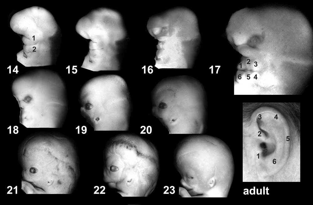
Images of the lateral view of the human embryonic head from week 5 (stage 14) through to week 8 (stage 23) showing development of the auricular hillocks that will form the external ear. The adult ear is also shown indicating the part of the ear that each hillock contributes.
Image Source: UNSW Embryology
File history
Click on a date/time to view the file as it appeared at that time.
| Date/Time | Thumbnail | Dimensions | User | Comment | |
|---|---|---|---|---|---|
| current | 23:57, 27 September 2009 | | 1,000 × 655 (42 KB) | S8600021 (talk | contribs) |
You cannot overwrite this file.
File usage
The following 23 pages use this file:
- 2009 Lecture 17
- 2010 Lecture 17
- 2011 Lab 10 - Late Embryo
- 2011 Lab 6 - Late Embryo
- AACP Meeting 2013 - Face Embryology
- ANAT2341 Lab 10 - Late Embryo
- ANAT2341 Lab 6 - Late Embryo
- Abnormal Development - Thalidomide
- BGDB Face and Ear - Late Embryo
- BGD Lecture - Face and Ear Development
- Carnegie stage 14
- Carnegie stage 15
- Carnegie stage 16
- Carnegie stage 17
- Carnegie stage 18
- Carnegie stage 19
- Carnegie stage 21
- Carnegie stage 22
- Carnegie stage 23
- Hearing - Outer Ear Development
- Kyoto Collection
- Lecture - Sensory Development
- Sensory - Hearing and Balance Development