File:External ear stages-14-23-adult.jpg: Difference between revisions
From Embryology
No edit summary |
|||
| Line 4: | Line 4: | ||
:'''Links:''' [[Hearing_-_Outer_Ear_Development]] | :'''Links:''' [[Hearing_-_Outer_Ear_Development]] | [[Carnegie_stage_14|Stage 14]] | [[Carnegie_stage_15|Stage 15]] | [[Carnegie_stage_16|Stage 16]] | [[Carnegie_stage_17|Stage 17]] | [[Carnegie_stage_18|Stage 18]] | [[Carnegie_stage_19|Stage 19]] | [[Carnegie_stage_20|Stage 20]] | [[Carnegie_stage_21|Stage 21]] | [[Carnegie_stage_22|Stage 22]] | [[Carnegie_stage_23|Stage 23]] | ||
Revision as of 12:01, 5 October 2011
External Ear
Images of the lateral view of the human embryonic head from week 5 (stage 14) through to week 8 (stage 23) showing development of the auricular hillocks that will form the external ear. The adult ear is also shown indicating the part of the ear that each hillock contributes.
- Links: Hearing_-_Outer_Ear_Development | Stage 14 | Stage 15 | Stage 16 | Stage 17 | Stage 18 | Stage 19 | Stage 20 | Stage 21 | Stage 22 | Stage 23
- Carnegie Stages: 1 | 2 | 3 | 4 | 5 | 6 | 7 | 8 | 9 | 10 | 11 | 12 | 13 | 14 | 15 | 16 | 17 | 18 | 19 | 20 | 21 | 22 | 23 | About Stages | Timeline
Image Source: UNSW Embryology
File history
Click on a date/time to view the file as it appeared at that time.
| Date/Time | Thumbnail | Dimensions | User | Comment | |
|---|---|---|---|---|---|
| current | 23:57, 27 September 2009 | 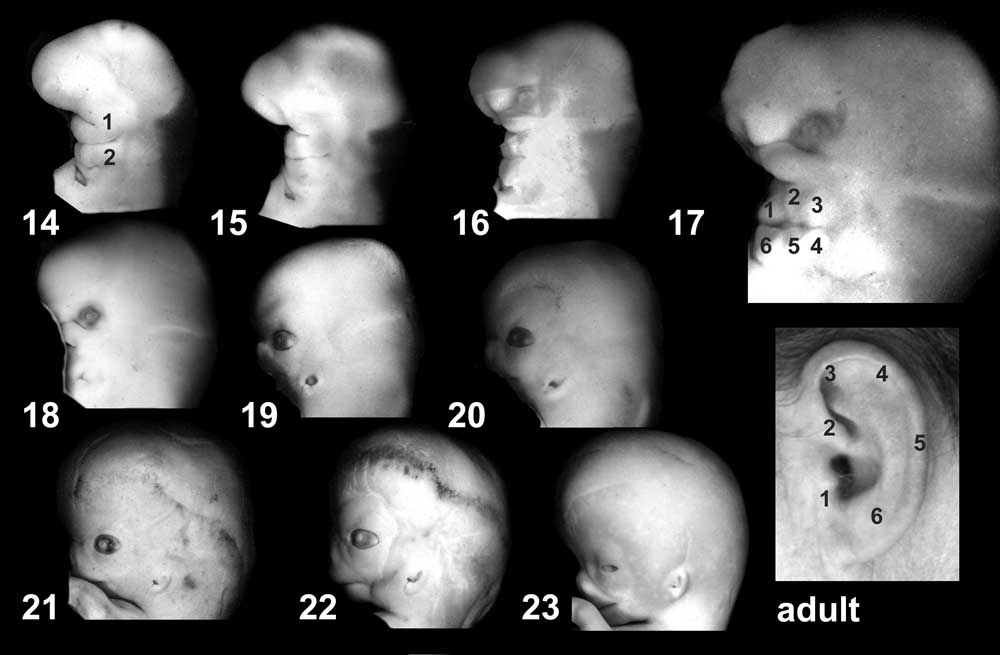 | 1,000 × 655 (42 KB) | S8600021 (talk | contribs) |
You cannot overwrite this file.
File usage
The following 23 pages use this file:
- 2009 Lecture 17
- 2010 Lecture 17
- 2011 Lab 10 - Late Embryo
- 2011 Lab 6 - Late Embryo
- AACP Meeting 2013 - Face Embryology
- ANAT2341 Lab 10 - Late Embryo
- ANAT2341 Lab 6 - Late Embryo
- Abnormal Development - Thalidomide
- BGDB Face and Ear - Late Embryo
- BGD Lecture - Face and Ear Development
- Carnegie stage 14
- Carnegie stage 15
- Carnegie stage 16
- Carnegie stage 17
- Carnegie stage 18
- Carnegie stage 19
- Carnegie stage 21
- Carnegie stage 22
- Carnegie stage 23
- Hearing - Outer Ear Development
- Kyoto Collection
- Lecture - Sensory Development
- Sensory - Hearing and Balance Development