File:Epithelium histology cartoon 01.jpg
From Embryology
No higher resolution available.
Epithelium_histology_cartoon_01.jpg (600 × 376 pixels, file size: 23 KB, MIME type: image/jpeg)
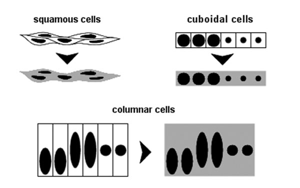
Epithelium Histology Cartoon
This simple cartoon shows the 3 main cellular shapes that are used to define epithelia: squamous, cuboidal, and columnar.
The image also shows the different histological appearances based upon the plane of sectioning.
Reference
Diagram prepared by Dr Carol Lazar.
Cite this page: Hill, M.A. (2024, April 23) Embryology Epithelium histology cartoon 01.jpg. Retrieved from https://embryology.med.unsw.edu.au/embryology/index.php/File:Epithelium_histology_cartoon_01.jpg
- © Dr Mark Hill 2024, UNSW Embryology ISBN: 978 0 7334 2609 4 - UNSW CRICOS Provider Code No. 00098G
File history
Click on a date/time to view the file as it appeared at that time.
| Date/Time | Thumbnail | Dimensions | User | Comment | |
|---|---|---|---|---|---|
| current | 16:04, 5 March 2012 | | 600 × 376 (23 KB) | Z8600021 (talk | contribs) |
You cannot overwrite this file.
File usage
The following 3 pages use this file: