File:Embryonic dorsal root ganglia in mouse.jpg: Difference between revisions
(Antibody stain against Neurofilament (green) and Ki 67 (red) in a Mouse embryo at day 12.5 after fertilization. Shown is the dorsal root ganglion (green ellipsoid regions where cells express neurofilament) and the ventricular zone (red region where cel...) |
No edit summary |
||
| Line 1: | Line 1: | ||
'''Fig. Embryonic dorsal root ganglia in mouse''' | |||
===Description=== | |||
Antibody stain against Neurofilament (green) and Ki 67 (red) in a Mouse embryo at day 12.5 after fertilization. Shown is the dorsal root ganglion (green ellipsoid regions where cells express neurofilament) and the ventricular zone (red region where cells proliferate) as well as the neural tube with roof and floor plate. | Antibody stain against Neurofilament (green) and Ki 67 (red) in a Mouse embryo at day 12.5 after fertilization. Shown is the dorsal root ganglion (green ellipsoid regions where cells express neurofilament) and the ventricular zone (red region where cells proliferate) as well as the neural tube with roof and floor plate. | ||
===Reference=== | |||
Creator: Hannes Röst (2007, May 4). Retrieved from https://commons.wikimedia.org/wiki/File:Mouse_NT_antibody_NF_Ki67.jpg | |||
===Copyright=== | |||
This file is released into the public domain, to which no exclusive intellectual property rights apply. This applies worldwide. In some countries this may not be legally possible; if so, the author grant anyone the right to use this work for any purpose, without any conditions, unless such conditions are required by law. | |||
{{Template:2018 Student Image}} | |||
Latest revision as of 18:54, 16 October 2018
Fig. Embryonic dorsal root ganglia in mouse
Description
Antibody stain against Neurofilament (green) and Ki 67 (red) in a Mouse embryo at day 12.5 after fertilization. Shown is the dorsal root ganglion (green ellipsoid regions where cells express neurofilament) and the ventricular zone (red region where cells proliferate) as well as the neural tube with roof and floor plate.
Reference
Creator: Hannes Röst (2007, May 4). Retrieved from https://commons.wikimedia.org/wiki/File:Mouse_NT_antibody_NF_Ki67.jpg
Copyright
This file is released into the public domain, to which no exclusive intellectual property rights apply. This applies worldwide. In some countries this may not be legally possible; if so, the author grant anyone the right to use this work for any purpose, without any conditions, unless such conditions are required by law.
- Note - This image was originally uploaded as part of an undergraduate science student 2018 project and may contain inaccuracies in either description or acknowledgements. Students have been advised in writing concerning the reuse of content and may accidentally have misunderstood the original terms of use. If image reuse on this non-commercial educational site infringes your existing copyright, please contact the site editor for immediate removal.
File history
Click on a date/time to view the file as it appeared at that time.
| Date/Time | Thumbnail | Dimensions | User | Comment | |
|---|---|---|---|---|---|
| current | 18:50, 16 October 2018 | 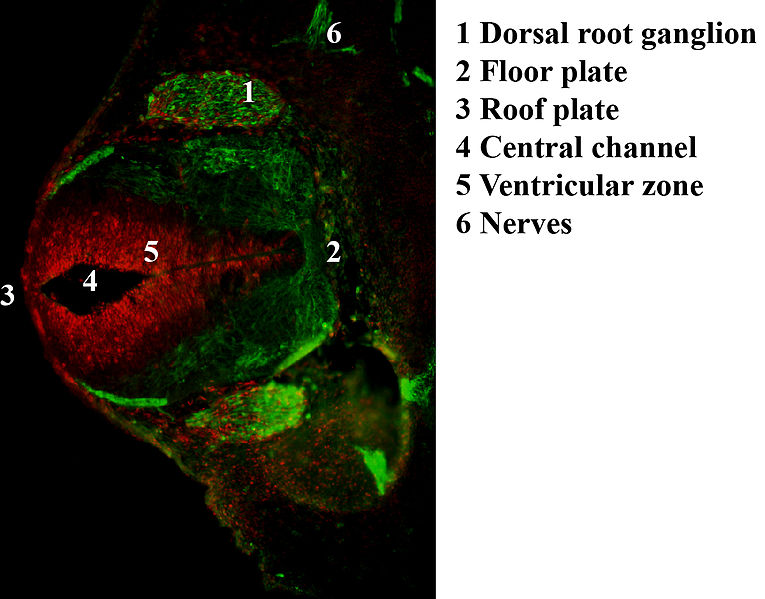 | 765 × 599 (84 KB) | Z5229431 (talk | contribs) | Antibody stain against Neurofilament (green) and Ki 67 (red) in a Mouse embryo at day 12.5 after fertilization. Shown is the dorsal root ganglion (green ellipsoid regions where cells express neurofilament) and the ventricular zone (red region where cel... |
You cannot overwrite this file.
File usage
The following 2 pages use this file: