File:Electrocardiograph findings in dogs affected with DMD.JPG
Electrocardiograph_findings_in_dogs_affected_with_DMD.JPG (609 × 538 pixels, file size: 64 KB, MIME type: image/jpeg)
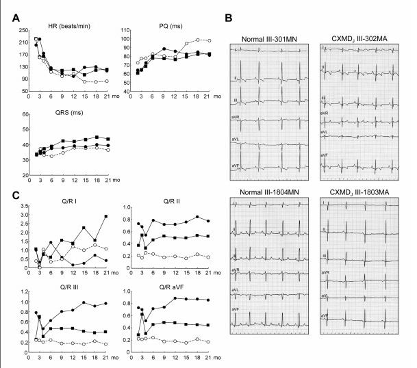
Electrocardiograph – device used to detect myocardial scarring. The graphs show an increased HR and a shortened PQ interval in DMD affected dogs.[1]
A: Sequential studies in heart rate (HR) (beats/min), PQ interval (ms) and duration (ms) of QRS complex. B: ECGs were recorded from normal dogs, III-301MN and III-1804MN, and CXMDJ dogs, III-302MA and III-1803MA, at 6 months of age. Distinct deep Q waves were present in the CXMDJ dogs. C: Sequential studies in Q/R ratios.
Reference
- ↑ Naoko Yugeta, Nobuyuki Urasawa, Yoko Fujii, Madoka Yoshimura, Katsutoshi Yuasa, Michiko R Wada et al. Cardiac involvement in Beagle-based canine X-linked muscular dystrophy in Japan (CXMDJ): electrocardiographic, echocardiographic, and morphologic studies.BMC Cardiovascular Disorders 2006, 6:47
This is an Open Access article distributed under the terms of the Creative Commons Attribution License (http://creativecommons.org/licenses/by/2.0), which permits unrestricted use, distribution, and reproduction in any medium, provided the original work is properly cited.
- Note - This image was originally uploaded as part of a student project and may contain inaccuracies in either description or acknowledgements. Students have been advised in writing concerning the reuse of content and may accidentally have misunderstood the original terms of use. If image reuse on this non-commercial educational site infringes your existing copyright, please contact the site editor for immediate removal.
Cite this page: Hill, M.A. (2024, April 25) Embryology Electrocardiograph findings in dogs affected with DMD.JPG. Retrieved from https://embryology.med.unsw.edu.au/embryology/index.php/File:Electrocardiograph_findings_in_dogs_affected_with_DMD.JPG
- © Dr Mark Hill 2024, UNSW Embryology ISBN: 978 0 7334 2609 4 - UNSW CRICOS Provider Code No. 00098G
File history
Click on a date/time to view the file as it appeared at that time.
| Date/Time | Thumbnail | Dimensions | User | Comment | |
|---|---|---|---|---|---|
| current | 12:54, 7 October 2011 | | 609 × 538 (64 KB) | Z3332327 (talk | contribs) | Electrocardiograph – device used to detect myocardial scarring. The graphs show an increased HR and a shortened PQ interval in DMD affected dogs.<ref>Naoko Yugeta, Nobuyuki Urasawa, Yoko Fujii, Madoka Yoshimura, Katsutoshi Yuasa, Michiko R Wada et al. C |
You cannot overwrite this file.
File usage
The following 2 pages use this file: