File:Ectopic pregnancy CT 03.jpg: Difference between revisions
No edit summary |
mNo edit summary |
||
| (2 intermediate revisions by 2 users not shown) | |||
| Line 7: | Line 7: | ||
* CT image demonstrates hematoma around uterus and hemoperitoneum. | * CT image demonstrates hematoma around uterus and hemoperitoneum. | ||
:'''Links:''' [[:File:Ectopic pregnancy CT 01.jpg|Ectopic pregnancy Initial CT]] | [[:File:Ectopic pregnancy CT 02.jpg|Ectopic pregnancy Follow-up CT]] | [[:File:Ectopic pregnancy CT 03.jpg|Ectopic pregnancy CT]] | [[Abnormal_Development_-_Ectopic_Implantation|Ectopic Implantation]] | [[Computed Tomography]] | |||
===Reference=== | ===Reference=== | ||
{{#pmid:20046504}} | |||
====Copyright==== | |||
This is an Open Access article distributed under the terms of the Creative Commons Attribution Non-Commercial License (http://creativecommons.org/licenses/by-nc/3.0) which permits unrestricted non-commercial use, distribution, and reproduction in any medium, provided the original work is properly cited. | This is an Open Access article distributed under the terms of the Creative Commons Attribution Non-Commercial License (http://creativecommons.org/licenses/by-nc/3.0) which permits unrestricted non-commercial use, distribution, and reproduction in any medium, provided the original work is properly cited. | ||
Original file name: Fig. 1 http://www.ncbi.nlm.nih.gov/pmc/articles/PMC2799642/figure/F1/ | |||
{{Footer}} | |||
[[Category:Ectopic Pregnancy]] [[Category:Abnormal Development]] [[Category:Computed Tomography]] | [[Category:Ectopic Pregnancy]] [[Category:Abnormal Development]] [[Category:Computed Tomography]] | ||
Latest revision as of 12:27, 1 June 2019
Ectopic Tubal Pregnancy Computed Tomography
Imaging findings are presented for 37-year-old woman with interstitial pregnancy.
- Coronal multiplanar reconstruction CT image shows heterogeneous enhancing mass (arrow) that abuts uterine fundus (arrowheads), suggestive of interstitial pregnancy.
- Note that there is hypodense line between mass and uterine fundus.
- CT image demonstrates hematoma around uterus and hemoperitoneum.
- Links: Ectopic pregnancy Initial CT | Ectopic pregnancy Follow-up CT | Ectopic pregnancy CT | Ectopic Implantation | Computed Tomography
Reference
Shin BS & Park MH. (2010). Incidental detection of interstitial pregnancy on CT imaging. Korean J Radiol , 11, 123-5. PMID: 20046504 DOI.
Copyright
This is an Open Access article distributed under the terms of the Creative Commons Attribution Non-Commercial License (http://creativecommons.org/licenses/by-nc/3.0) which permits unrestricted non-commercial use, distribution, and reproduction in any medium, provided the original work is properly cited.
Original file name: Fig. 1 http://www.ncbi.nlm.nih.gov/pmc/articles/PMC2799642/figure/F1/
Cite this page: Hill, M.A. (2024, April 19) Embryology Ectopic pregnancy CT 03.jpg. Retrieved from https://embryology.med.unsw.edu.au/embryology/index.php/File:Ectopic_pregnancy_CT_03.jpg
- © Dr Mark Hill 2024, UNSW Embryology ISBN: 978 0 7334 2609 4 - UNSW CRICOS Provider Code No. 00098G
File history
Click on a date/time to view the file as it appeared at that time.
| Date/Time | Thumbnail | Dimensions | User | Comment | |
|---|---|---|---|---|---|
| current | 15:02, 3 September 2011 | 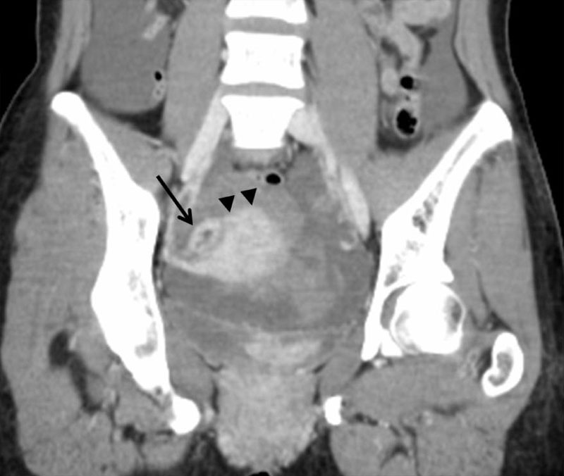 | 800 × 676 (54 KB) | S8600021 (talk | contribs) | Ectopic---PMC2799642-C.jpg |
You cannot overwrite this file.
File usage
The following 2 pages use this file: