File:Early zygote.jpg: Difference between revisions
From Embryology
No edit summary |
|||
| Line 6: | Line 6: | ||
* there would still be granulosa cells and spermatozoa attached to the zone pellucida. | * there would still be granulosa cells and spermatozoa attached to the zone pellucida. | ||
* the zygote floats freely within the uterine tube. | * the zygote floats freely within the uterine tube. | ||
* The cell is preparing for the first mitotic division. | |||
Revision as of 12:01, 10 March 2012
Early Zygote
This is described as an early human zygote as the male and female pronuclei have not yet combined to form a single nucleus.
At this stage in vivo:
- there would still be granulosa cells and spermatozoa attached to the zone pellucida.
- the zygote floats freely within the uterine tube.
- The cell is preparing for the first mitotic division.
About Carnegie Stages 1
Facts: Week 1, size 0.1 - 0.15 mm (100 - 150 microns)
Features: zygote, fertilized oocyte, pronuclei, polar bodies, zona pellucida
Image Source: UNSW Embryology http://embryology.med.unsw.edu.au/wwwhuman/Stages/Stage1.htm
File history
Click on a date/time to view the file as it appeared at that time.
| Date/Time | Thumbnail | Dimensions | User | Comment | |
|---|---|---|---|---|---|
| current | 11:45, 10 March 2012 |  | 500 × 441 (23 KB) | Z8600021 (talk | contribs) | |
| 16:40, 27 July 2009 | 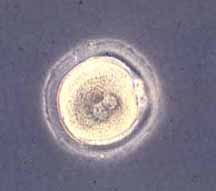 | 216 × 191 (4 KB) | MarkHill (talk | contribs) | About Carnegie Stages 1 Facts: Week 1, size 0.1-0.15 mm Features: zygote, fertilized oocyte, pronuclei, polar bodies, zona pellucida Image Source: UNSW Embryology http://embryology.med.unsw.edu.au/wwwhuman/Stages/Stage1.htm |
You cannot overwrite this file.
File usage
The following 88 pages use this file:
- 2009 Lab 12
- 2009 Lecture 2
- 2009 Lecture 3
- 2010 BGD Lecture - Development of the Embryo/Fetus 1
- 2010 BGD Lecture - Development of the Embryo/Fetus 2
- 2010 BGD Practical 3 - Early Cell Division
- 2010 BGD Practical 3 - Week 3 Summary
- 2010 BGD Practical 6 - Week 8
- 2010 BGD Tutorial - Applied Embryology and Teratology
- 2010 Foundations Lecture - Introduction to Human Development
- 2010 Foundations Practical - Introduction to Human Development
- 2010 Lab 2
- 2010 Lecture 2
- 2010 Lecture 3
- 2010 Lecture 6
- 2011 Group Project 11
- ANAT2341Embryology Image Tutorial
- ANAT2341 Embryology 2015
- ANAT2341 Embryology 2016
- ANAT2341 Lab 1 - Fertilization
- ANAT2341 Lab 2 - Week 1
- ANZACA Meeting 2012 - Embryology
- Abnormal Development - Environmental
- Abnormal Development - Illegal Drugs
- Abnormal Development - Teratogens
- Abnormal Development - Twinning
- BGDA Lecture - Development of the Embryo/Fetus 2
- BGDA Practical 3 - Fertilization
- BGDA Practical 3 - Week 1 Summary
- BGDA Practical 3 - Week 2 Summary
- BGDA Practical 3 - Week 3 Summary
- BGDA Practical 7 - Week 8
- BGD Tutorial - Applied Embryology and Teratology
- Carnegie stage 1
- Editing Basics
- Embryology Image Tutorial
- Embryonic Development
- Fertilization
- Foundations Lecture - Introduction to Human Development
- Foundations Practical - Critical Periods
- Foundations Practical - Introduction to Human Development
- Foundations Practical - Week 1 and 2
- Human Abnormal Development
- Lecture - 2012 Course Introduction
- Lecture - 2013 Course Introduction
- Lecture - Ectoderm Development
- Lecture - Fetal Development
- Lecture - Week 1 and 2 Development
- Placenta - Membranes
- Pre-Medicine Program - Embryology
- Test Student 2010
- Timeline human development
- UNSW Learning and Teaching Seminar 2012
- Week 1
- Week 1 - Abnormalities
- Week 2
- Zona pellucida
- Talk:2010 BGD Lecture - Development of the Embryo/Fetus 2
- Talk:Timeline human development
- User:Z3186755
- User:Z3216889
- User:Z3224500
- User:Z3241780
- User:Z3252083
- User:Z3252635
- User:Z3252833
- User:Z3254433
- User:Z3254753
- User:Z3265772
- User:Z3288088
- User:Z3290040
- User:Z3291079
- User:Z3292208
- User:Z3292955
- User:Z3293029
- User:Z3305561
- User:Z3318446
- User:Z5163485
- File:Human-critical periods of development.jpg
- File talk:Human-critical periods.jpg
- Template:Critical Periods table
- Template:First Trimester Timeline
- Template:First Trimester Timeline collapsable table
- Template:Monoygotic Twinning Table
- Template:Week 1 table
- Template talk:First Trimester Timeline
- Help:Editing Basics
- Help:Image Tutorial