File:Dystrophin in the muscle fibre membrane.jpg
Original file (1,840 × 1,355 pixels, file size: 415 KB, MIME type: image/jpeg)
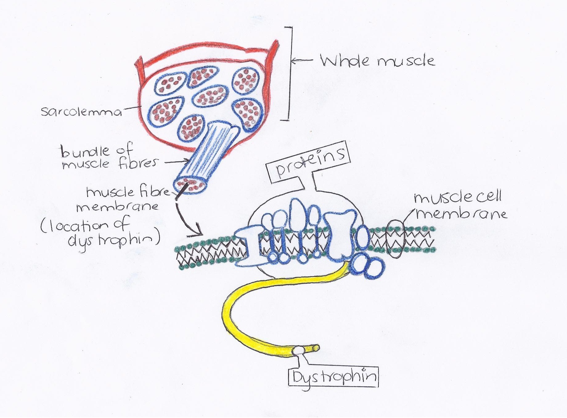
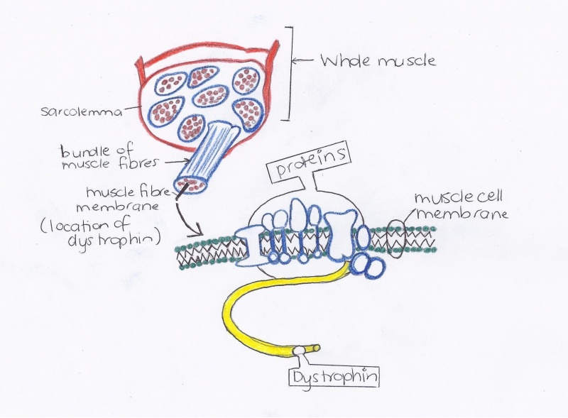
This image is a visual display of the muscle fibre membrane and the location of dystrophin within the sarcolemma surrounding each muscle fibre. There are 4 main transmembrane proteins that compose the dystrophin-glycoprotein complex (DGP), also referred to as the dystrophin-associated protein complex (DAPC), located in the muscle fibre membrane. These proteins include:
- Sarcoglycans: a family of transmembrane proteins that work in conjunction with other proteins (namely dystrophin) to protect muscle fibres during contraction. There are 4 main types present in striated muscle, including: α-sarcoglycan, β-sarcoglycan, γ-sarcoglycan and δ-sarcoglycan. The sarcoglycan complex is disrupted and destabilized from the plasma membrane when dystrophin is mutated and hence leads to eccentric contraction-induced disruption of the plasma membrane of smooth and cardiac muscle. [1]
- Dystroglycan: connects the extracellular matrix and cytoskeleton and also forms a linkage between the dystrophin-glycoprotein complex and the basal lamina. When dystrophin is mutated, these linkages formed by dystroglycan are lost and also cause the disruption of the plasma membrane.[2]
- Syntrophin: links to the extracellular matrix through dystrophin and creates signal transduction complexes at the DAPC and therefore when dystrophin is mutated, this crucial signalling pathway is lost. [3]
- Dystrobrevinis: the functional role of this complex has not yet been clearly identified, however studies show that dystrobrevin disappears from the muscle membrane in Duchenne muscular dystrophy (DMD). [4]
Image creator: Ashleigh Pontifex (z3332629)
Student image constructed based on the image presented on: http://www.mda.org/publications/fa-dmdbmd-what.html
Location of dystrophin within the muscle fibre membrane by Ashleigh Pontifex is licensed under a Creative Commons Attribution-NoDerivs 3.0 Unported License. Based on a work at www.mda.org.
<a rel="license" href="http://creativecommons.org/licenses/by-nd/3.0/"><img alt="Creative Commons License" style="border-width:0" src="http://i.creativecommons.org/l/by-nd/3.0/88x31.png" /></a>
Location of dystrophin within the muscle fibre membrane by Ashleigh Pontifex is licensed under a <a rel="license" href="http://creativecommons.org/licenses/by-nd/3.0/">Creative Commons Attribution-NoDerivs 3.0 Unported License</a>.
Based on a work at <a xmlns:dct="http://purl.org/dc/terms/" href="http://www.mda.org/publications/fa-dmdbmd-what.html" rel="dct:source">www.mda.org</a>.
- Note - This image was originally uploaded as part of a student project and may contain inaccuracies in either description or acknowledgements. Students have been advised in writing concerning the reuse of content and may accidentally have misunderstood the original terms of use. If image reuse on this non-commercial educational site infringes your existing copyright, please contact the site editor for immediate removal.
Cite this page: Hill, M.A. (2024, April 19) Embryology Dystrophin in the muscle fibre membrane.jpg. Retrieved from https://embryology.med.unsw.edu.au/embryology/index.php/File:Dystrophin_in_the_muscle_fibre_membrane.jpg
- © Dr Mark Hill 2024, UNSW Embryology ISBN: 978 0 7334 2609 4 - UNSW CRICOS Provider Code No. 00098G.
- ↑ Hack, A.A., Lam, M, J.,Cordier, L., Shoturma, D.I (2000). “Differential requirement for individual sarcoglycans and dystrophin in the assembly and function of the dystrophin-glycoprotein complex”. Journal of Cell Science 113, page 2535-2544 (2000). Accessed via: http://jcs.biologists.org/content/113/14/2535.full.pdf
- ↑ Neuromuscular (2011). “Membrane-associated protein complexes in skeletal muscle fibres and connective tissue.” Author anonymous. Accessed via: http://neuromuscular.wustl.edu/musdist/dag2.htm#ad
- ↑ Davies.K.E, Nowak. K.J (2006). "Molecular mechanisms of musclar dystrophies: old and new players." Nature Reviews. October, 2006 Volume 7, pages 763-773.
- ↑ Grisoni,K., Gieseler,K., Se´ galat,L. (2002). “Dystrobrevin requires a dystrophin-binding domain to function in Caenorhabditis elegans”. Eur. J. Biochem. 269, pages 1607±1612.
File history
Click on a date/time to view the file as it appeared at that time.
| Date/Time | Thumbnail | Dimensions | User | Comment | |
|---|---|---|---|---|---|
| current | 08:58, 4 October 2011 | | 1,840 × 1,355 (415 KB) | Z3332629 (talk | contribs) | This image is a visual display of the muscle fibre membrane and the location of dystrophin within the sarcolemma surrounding each muscle fibre. {{Template:2011 Student Image}}. Image creator: Ashleigh Pontifex (z3332629) Student image constructed bas |
You cannot overwrite this file.
File usage
There are no pages that use this file.