File:Drawing Of A Normal Heart.PNG: Difference between revisions
No edit summary |
No edit summary |
||
| Line 1: | Line 1: | ||
Figure name: The normal heart | Figure name: The four features of the tetralogy of Fallot in comparison to the normal heart.(A) '''normal heart''', | ||
(B) ventricular septal defects, (C) obstruction of right ventricular outflow, (D) "Overriding" aorta, (E) Right | |||
ventricular hypertrophy. Blue: deoxygenated blood; red: oxygenated blood; RA= right atrium; RV= right | |||
ventricle; LA= left atrium; LV= left ventricle; Ao= Arch of aorta; direction of blood flow. | ventricle; LA= left atrium; LV= left ventricle; Ao= Arch of aorta; direction of blood flow. | ||
While this drawings illustrate of each feature in isolated state for better understanding. However, it is | |||
important to note, that the features shown in A-E commonly occur together. | |||
Drawings by Anna Marx based on Robbins Basic Pathology <ref> Kumar, V., Abbas, A., Fausto, N., Mitchell, R. N. (2007). Robbins Basic Pathology. In Saunders Elsevier (Ed 8), Philadelphia. https://evolve.elsevier.com/productPages/s_1221.html</ref> <ref><pubmed>18636635</pubmed></ref>. | Drawings by Anna Marx based on Robbins Basic Pathology <ref> Kumar, V., Abbas, A., Fausto, N., Mitchell, R. N. (2007). Robbins Basic Pathology. In Saunders Elsevier (Ed 8), Philadelphia. https://evolve.elsevier.com/productPages/s_1221.html</ref> <ref><pubmed>18636635</pubmed></ref>. | ||
Latest revision as of 11:29, 15 September 2011
Figure name: The four features of the tetralogy of Fallot in comparison to the normal heart.(A) normal heart, (B) ventricular septal defects, (C) obstruction of right ventricular outflow, (D) "Overriding" aorta, (E) Right ventricular hypertrophy. Blue: deoxygenated blood; red: oxygenated blood; RA= right atrium; RV= right ventricle; LA= left atrium; LV= left ventricle; Ao= Arch of aorta; direction of blood flow.
While this drawings illustrate of each feature in isolated state for better understanding. However, it is important to note, that the features shown in A-E commonly occur together.
Drawings by Anna Marx based on Robbins Basic Pathology [1] [2].
- ↑ Kumar, V., Abbas, A., Fausto, N., Mitchell, R. N. (2007). Robbins Basic Pathology. In Saunders Elsevier (Ed 8), Philadelphia. https://evolve.elsevier.com/productPages/s_1221.html
- ↑ <pubmed>18636635</pubmed>
File history
Click on a date/time to view the file as it appeared at that time.
| Date/Time | Thumbnail | Dimensions | User | Comment | |
|---|---|---|---|---|---|
| current | 11:02, 15 September 2011 | 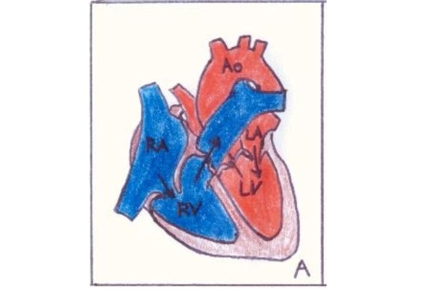 | 840 × 566 (409 KB) | Z3288729 (talk | contribs) | Image Drawn by Anna Marx |
You cannot overwrite this file.
File usage
The following 2 pages use this file: