File:Drawing Of A Normal Heart.PNG: Difference between revisions
From Embryology
(Image Drawn by Anna Marx) |
No edit summary |
||
| Line 1: | Line 1: | ||
Figure name: The normal heart | |||
Legend: Blue: deoxygenated blood; red: oxygenated blood; RA= right atrium; RV= right | |||
ventricle; LA= left atrium; LV= left ventricle; Ao= Arch of aorta; direction of blood flow. | |||
Compare this drawing of the normal heart with each of the individual features of tetralogy of fallot shown in the images B to E. | |||
Drawings by Anna Marx based on Robbins Basic Pathology <ref> Kumar, V., Abbas, A., Fausto, N., Mitchell, R. N. (2007). Robbins Basic Pathology. In Saunders Elsevier (Ed 8), Philadelphia. https://evolve.elsevier.com/productPages/s_1221.html</ref> <ref><pubmed>18636635</pubmed></ref>. | |||
Revision as of 11:26, 15 September 2011
Figure name: The normal heart Legend: Blue: deoxygenated blood; red: oxygenated blood; RA= right atrium; RV= right ventricle; LA= left atrium; LV= left ventricle; Ao= Arch of aorta; direction of blood flow.
Compare this drawing of the normal heart with each of the individual features of tetralogy of fallot shown in the images B to E.
Drawings by Anna Marx based on Robbins Basic Pathology [1] [2].
- ↑ Kumar, V., Abbas, A., Fausto, N., Mitchell, R. N. (2007). Robbins Basic Pathology. In Saunders Elsevier (Ed 8), Philadelphia. https://evolve.elsevier.com/productPages/s_1221.html
- ↑ <pubmed>18636635</pubmed>
File history
Click on a date/time to view the file as it appeared at that time.
| Date/Time | Thumbnail | Dimensions | User | Comment | |
|---|---|---|---|---|---|
| current | 11:02, 15 September 2011 | 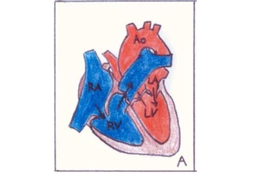 | 840 × 566 (409 KB) | Z3288729 (talk | contribs) | Image Drawn by Anna Marx |
You cannot overwrite this file.
File usage
The following 2 pages use this file: