File:Double Outlet Right Ventricle.jpg: Difference between revisions
From Embryology
| Line 5: | Line 5: | ||
* Arrangement of the atrioventricular valves and the ventriculoarterial connections are variable. | * Arrangement of the atrioventricular valves and the ventriculoarterial connections are variable. | ||
* Clinical manifestations variable. | * Clinical manifestations variable. | ||
{{Template:Heart Links}} | |||
{{Template:Manitoba Health Report 2001}} | {{Template:Manitoba Health Report 2001}} | ||
[[Category:Cardiovascular]] [[Category:Heart]] [[Category:Abnormal Development]] | [[Category:Cardiovascular]] [[Category:Heart]] [[Category:Abnormal Development]] | ||
Revision as of 10:29, 2 August 2012
Heart Abnormality - Double Outlet Right Ventricle
- 1-1.5% of Congenital Heart Disease
- Both large arteries arise wholly or mainly from the right ventricle.
- Arrangement of the atrioventricular valves and the ventriculoarterial connections are variable.
- Clinical manifestations variable.
| Heart Abnormal: Tutorial Abnormalities | atrial septal defects | double outlet right ventricle | hypoplastic left heart | patent ductus arteriosus | transposition of the great vessels | Tetralogy of Fallot | ventricular septal defects | coarctation of the aorta | Category ASD | Category PDA | Category ToF | Category VSD | ICD10 - Cardiovascular | ICD11 |
Image source: Report of the Review and Implementation Committee for The Report of the Manitoba Pediatric Cardiac Surgery Inquest May 2001 image showing heart abnormalities courtesy Government of Manitoba.
File history
Click on a date/time to view the file as it appeared at that time.
| Date/Time | Thumbnail | Dimensions | User | Comment | |
|---|---|---|---|---|---|
| current | 11:23, 14 March 2010 | 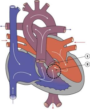 | 289 × 350 (16 KB) | Z3212774 (talk | contribs) | category:Heart ILP |
You cannot overwrite this file.
File usage
The following 12 pages use this file:
- ANAT2341 Lab 11 - Heart Abnormalities
- Advanced - Abnormalities
- Cardiovascular System - Abnormalities
- Cardiovascular System - Coarctation of the Aorta
- Cardiovascular System - Double Outlet Right Ventricle
- Fetal ECHO Meeting 2012
- Intermediate - Cardiac Abnormalities
- RPAH Cardiac Embryology 2014
- Talk:Cardiovascular System - Abnormalities
- Template:Heart Abnormality Cartoons
- Template:Heart abnormal cartoon gallery
- Template talk:Heart abnormal cartoon gallery