File:Different Stages of Embryo Development.jpeg: Difference between revisions
(S12978-015-0031-x-2.jpg PMID 25935518) |
No edit summary |
||
| Line 1: | Line 1: | ||
==Different Stages of Embryo Development== | |||
PMID 25935518 | Different stages of embryo development captured with a Time Lapse System. The images were captured (Embrioscope, Fertilitech-Unisence Denmark, Embryo-Viewer Software) at different stages of the embryo’s development post injection: a – 2 pro-nucleai, 16 hours, b – 2 cells (30 hours), c – 4 cells (39 hours), d – 8 cells (65 hours), e – start of blastocyst (88 hours), f – expanded blastocyst (99 hour), g – hatching blastocyst (106 hours). | ||
(Original figure legend, image based on data from Doroftei et al. PMID 25935518) | |||
===Reference=== | |||
<pubmed>25935518</pubmed>| [http://www.reproductive-health-journal.com/content/12/1/38] | |||
====Copyright==== | |||
© 2015 Doroftei et al.; licensee BioMed Central. | |||
This is an Open Access article distributed under the terms of the Creative Commons Attribution License (http://creativecommons.org/licenses/by/4.0), which permits unrestricted use, distribution, and reproduction in any medium, provided the original work is properly credited. The Creative Commons Public Domain Dedication waiver (http://creativecommons.org/publicdomain/zero/1.0/) applies to the data made available in this article, unless otherwise stated. | |||
Figure 2: S12978-015-0031-x-2.jpg | |||
{{Template:Student Image}} | |||
Latest revision as of 18:13, 14 August 2015
Different Stages of Embryo Development
Different stages of embryo development captured with a Time Lapse System. The images were captured (Embrioscope, Fertilitech-Unisence Denmark, Embryo-Viewer Software) at different stages of the embryo’s development post injection: a – 2 pro-nucleai, 16 hours, b – 2 cells (30 hours), c – 4 cells (39 hours), d – 8 cells (65 hours), e – start of blastocyst (88 hours), f – expanded blastocyst (99 hour), g – hatching blastocyst (106 hours).
(Original figure legend, image based on data from Doroftei et al. PMID 25935518)
Reference
<pubmed>25935518</pubmed>| [1]
Copyright
© 2015 Doroftei et al.; licensee BioMed Central. This is an Open Access article distributed under the terms of the Creative Commons Attribution License (http://creativecommons.org/licenses/by/4.0), which permits unrestricted use, distribution, and reproduction in any medium, provided the original work is properly credited. The Creative Commons Public Domain Dedication waiver (http://creativecommons.org/publicdomain/zero/1.0/) applies to the data made available in this article, unless otherwise stated.
Figure 2: S12978-015-0031-x-2.jpg
- Note - This image was originally uploaded as part of an undergraduate science student project and may contain inaccuracies in either description or acknowledgements. Students have been advised in writing concerning the reuse of content and may accidentally have misunderstood the original terms of use. If image reuse on this non-commercial educational site infringes your existing copyright, please contact the site editor for immediate removal.
File history
Click on a date/time to view the file as it appeared at that time.
| Date/Time | Thumbnail | Dimensions | User | Comment | |
|---|---|---|---|---|---|
| current | 18:08, 14 August 2015 | 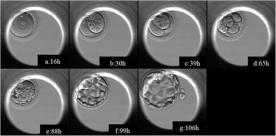 | 567 × 279 (36 KB) | Z5088434 (talk | contribs) | S12978-015-0031-x-2.jpg PMID 25935518 |
You cannot overwrite this file.
File usage
The following page uses this file: