File:Development of Trilaminar Embryonic Disc .png: Difference between revisions
No edit summary |
No edit summary |
||
| Line 1: | Line 1: | ||
==Description== | ==Description== | ||
| Line 15: | Line 10: | ||
(5) Fusion of the bilateral vitelline veins generates the primary heart tube. The point of fusion is recognized as the ventral mesocardium. The medial splanchnic mesoderm becomes the pericardial back wall, which is connected to the heart tube via the dorsal mesocardium. | (5) Fusion of the bilateral vitelline veins generates the primary heart tube. The point of fusion is recognized as the ventral mesocardium. The medial splanchnic mesoderm becomes the pericardial back wall, which is connected to the heart tube via the dorsal mesocardium. | ||
(6) Ventral view of the primary heart tube. The height of cross sections (4) and (5) is indicated by arrows | (6) Ventral view of the primary heart tube. The height of cross sections (4) and (5) is indicated by arrows | ||
===Reference=== | |||
[https://doi.org/10.1371/journal.pone.0022055.g001] | |||
===Copyright=== | |||
Open access, Peer Reviewed | |||
{{Template:Student Image}} | |||
Revision as of 23:12, 17 September 2017
Description
Figure 1
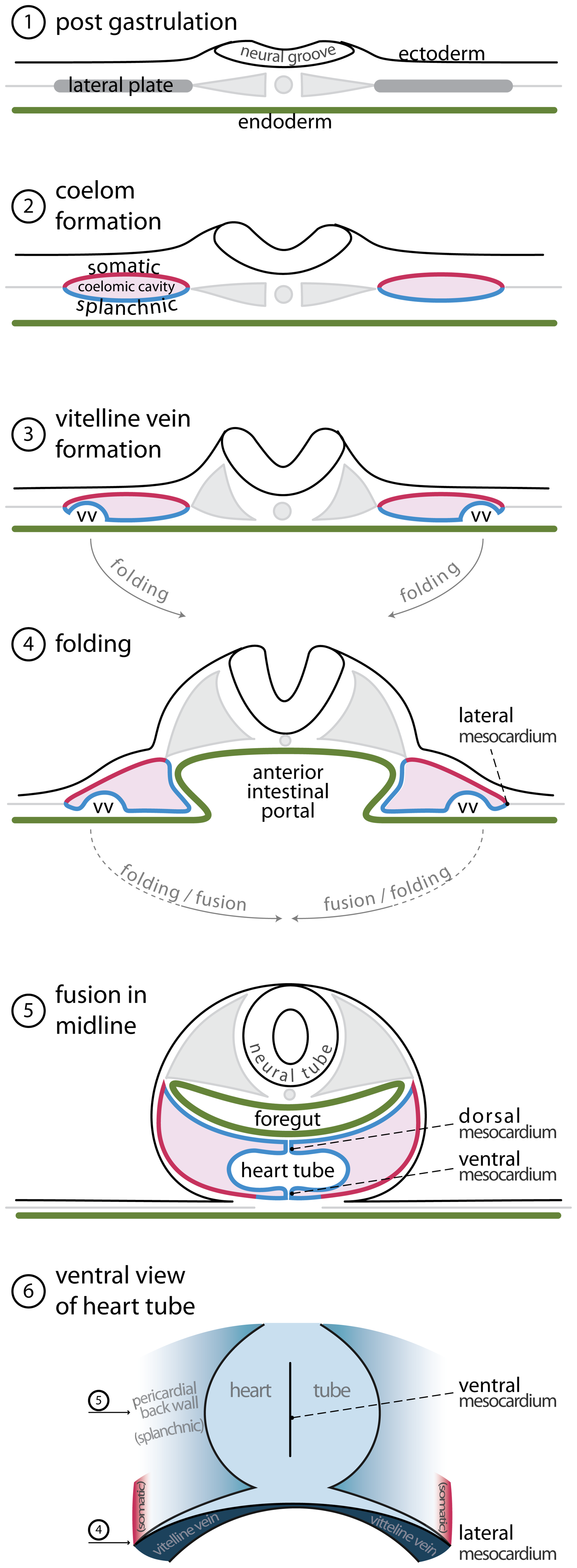
(1) Trilaminalar embryonic disc after gastrulation; lateral plate mesoderm resides between the endoderm (green) and the ectoderm (black). (2) The lateral plate mesoderm separates in a splanchnic (blue) and a somatic (red) layer by the formation of the coelomic cavity (violet). (3) The lateral edges of the splanchnic mesoderm luminize, forming the bilateral vitelline veins. (4) By folding the lateral edges of the embryonic disc move inwards, creating the anterior intestinal portal. The persisting contact between the somatic and splanchnic mesoderm is called the lateral mesocardium. (5) Fusion of the bilateral vitelline veins generates the primary heart tube. The point of fusion is recognized as the ventral mesocardium. The medial splanchnic mesoderm becomes the pericardial back wall, which is connected to the heart tube via the dorsal mesocardium. (6) Ventral view of the primary heart tube. The height of cross sections (4) and (5) is indicated by arrows
Reference
Copyright
Open access, Peer Reviewed
- Note - This image was originally uploaded as part of an undergraduate science student project and may contain inaccuracies in either description or acknowledgements. Students have been advised in writing concerning the reuse of content and may accidentally have misunderstood the original terms of use. If image reuse on this non-commercial educational site infringes your existing copyright, please contact the site editor for immediate removal.
File history
Click on a date/time to view the file as it appeared at that time.
| Date/Time | Thumbnail | Dimensions | User | Comment | |
|---|---|---|---|---|---|
| current | 22:08, 17 September 2017 | 1,921 × 5,223 (1.24 MB) | Z5076019 (talk | contribs) | [https://doi.org/10.1371/journal.pone.0022055.g001] Open access, Peer Reviewed |
You cannot overwrite this file.
File usage
There are no pages that use this file.