File:Developing vertebra.jpg
From Embryology
No higher resolution available.
Developing_vertebra.jpg (558 × 428 pixels, file size: 93 KB, MIME type: image/jpeg)
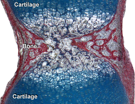
Cross-section of developing vertebra (longitudinal of axial skeleton).
Centre of the image shows initial vertebra "bony collar" (in red) forming, either side of which is the original cartilage (blue) "template", outside of that again are the 2 intervertebral discs (not shown).
Source: UNSW Embryology http://embryology.med.unsw.edu.au/Notes/skmus8.htm#Histology
File history
Click on a date/time to view the file as it appeared at that time.
| Date/Time | Thumbnail | Dimensions | User | Comment | |
|---|---|---|---|---|---|
| current | 10:44, 11 September 2009 | | 558 × 428 (93 KB) | S8600021 (talk | contribs) | Cross-section of developing vertebra (longitudinal of axial skeleton). Centre of the image shows initial vertebra "bony collar" (in red) forming, either side of which is the original cartilage (blue) "template", outside of that again are the 2 intervert |
You cannot overwrite this file.
File usage
The following 10 pages use this file:
- 2009 Lecture 13
- 2010 Lecture 13
- Lecture - Musculoskeletal Development
- Musculoskeletal System - Abnormalities
- Musculoskeletal System - Axial Skeleton Development
- Musculoskeletal System - Bone Development
- Musculoskeletal System - Cartilage Development
- Musculoskeletal System - Joint Development
- Musculoskeletal System - Pelvis Development
- Musculoskeletal System Development