File:Developing tongue histology 001.jpg
From Embryology
Size of this preview: 776 × 599 pixels. Other resolution: 900 × 695 pixels.
Original file (900 × 695 pixels, file size: 334 KB, MIME type: image/jpeg)
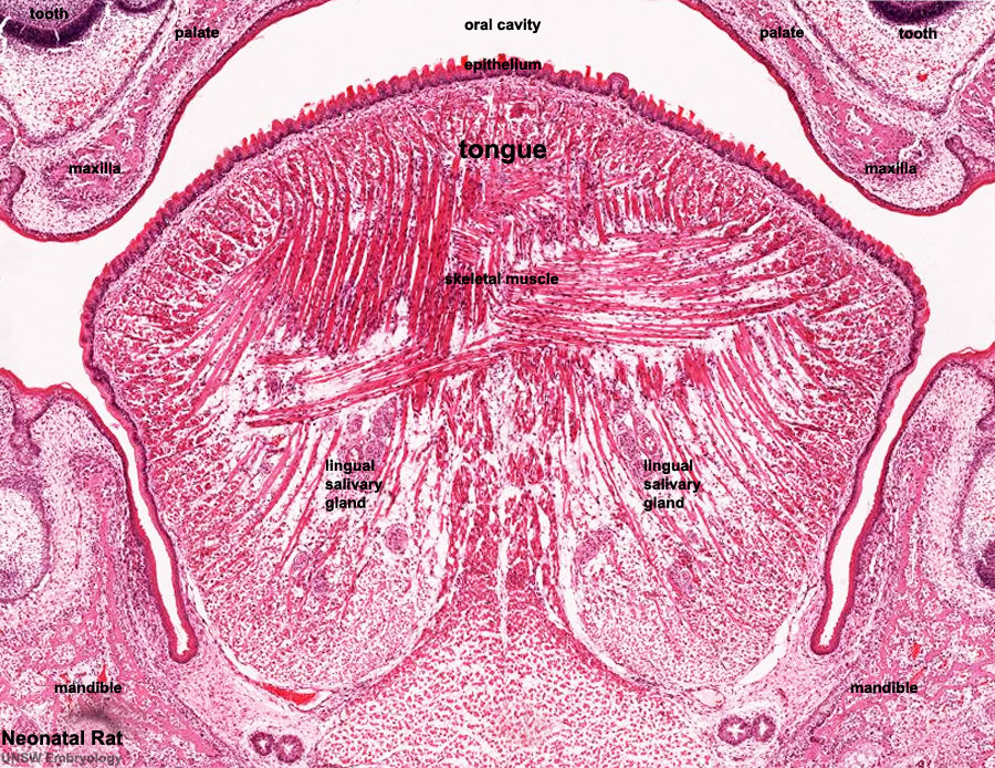
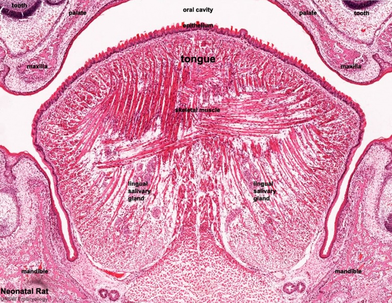
Neonatal Rat Tongue Histology
Section through the newborn rat oral cavity. Note the different orientations of the developing muscle fibres. (Stain - Haematoxylin Eosin)
- developing tongue epithelium
- developing tongue skeletal muscle
- lingual salivary glands
- Palate
- Maxilla
- Mandible
- Links: Tongue Development
Cite this page: Hill, M.A. (2024, April 20) Embryology Developing tongue histology 001.jpg. Retrieved from https://embryology.med.unsw.edu.au/embryology/index.php/File:Developing_tongue_histology_001.jpg
- © Dr Mark Hill 2024, UNSW Embryology ISBN: 978 0 7334 2609 4 - UNSW CRICOS Provider Code No. 00098G
File history
Click on a date/time to view the file as it appeared at that time.
| Date/Time | Thumbnail | Dimensions | User | Comment | |
|---|---|---|---|---|---|
| current | 00:27, 4 October 2011 | | 900 × 695 (334 KB) | S8600021 (talk | contribs) | ==Neonatal Rat Tongue Histology== * Stain HE Category:Rat Category:Tongue |
You cannot overwrite this file.
File usage
The following 3 pages use this file: