File:Day 16 Reposition of umbilical hernia.JPG

Day_16_Reposition_of_umbilical_hernia.JPG (640 × 532 pixels, file size: 34 KB, MIME type: image/jpeg)
Day 16: the umbilical hernia repositions. superficial veins are visible through the transparet skin of the embryo.
Vc= vena cephalica, Vth= Vena thoracica lateralis, Vt= Vena temporalis superficialis, Vu= Vena umbilica, Vv= Vena vitellina, P= Placenta, Vm+ Vena metatarseae dorsales, Nh= umbilical hernia
Illustration by z3252340, based upon Fig 345 and Fig 246, in Dr Karl Theiler’s ‘The House Mouse; Atlas of Embryonic Development’, Springer - Verlag New York Inc, New York, 1989
Beginning six months after publication, I, z3252340, grant the public the non-exclusive right to copy, distribute, or display the Work under a Creative Commons Attribution-Noncommercial-Share Alike 3.0 Unported license, as described at http://creativecommons.org/licenses/by-nc-sa/3.0/ and http://creativecommons.org/licenses/by-nc-sa/3.0/legalcode.
File history
Click on a date/time to view the file as it appeared at that time.
| Date/Time | Thumbnail | Dimensions | User | Comment | |
|---|---|---|---|---|---|
| current | 14:07, 19 September 2009 | | 640 × 532 (34 KB) | Z3252340 (talk | contribs) | Reverted to version as of 04:06, 19 September 2009 |
| 14:06, 19 September 2009 | 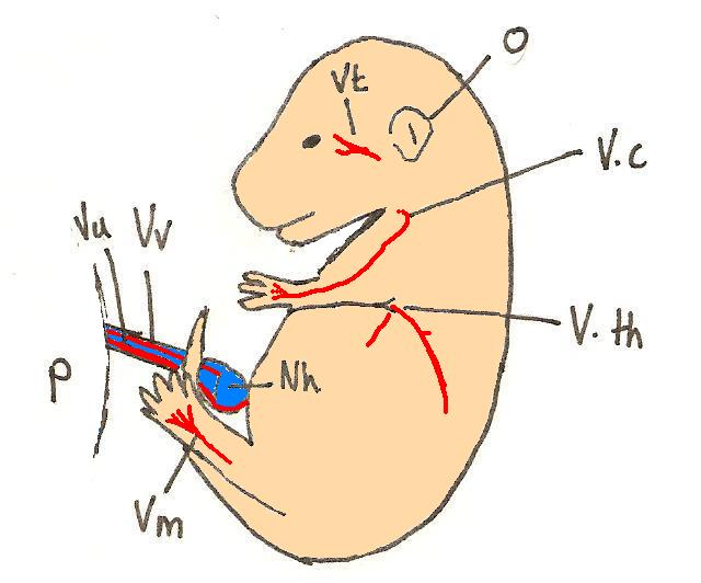 | 640 × 532 (34 KB) | Z3252340 (talk | contribs) | Reverted to version as of 04:05, 19 September 2009 | |
| 14:06, 19 September 2009 |  | 640 × 532 (34 KB) | Z3252340 (talk | contribs) | ||
| 14:05, 19 September 2009 |  | 640 × 532 (34 KB) | Z3252340 (talk | contribs) | colour | |
| 10:44, 16 September 2009 | 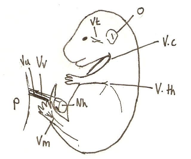 | 640 × 532 (33 KB) | Z3252340 (talk | contribs) | Day 16: the umbilical hernia repositions. superficial veins are visible through the transparet skin of the embryo. Vc= vena cephalica, Vth= Vena thoracica lateralis, Vt= Vena temporalis superficialis, Vu= Vena umbilica, Vv= Vena vitellina, P= Placenta, |
You cannot overwrite this file.
File usage
The following 3 pages use this file: