File:Day 11.5 Lens Vesicle completely separated from surface.JPG: Difference between revisions
No edit summary |
mNo edit summary |
||
| Line 6: | Line 6: | ||
[[ | :'''Links:''' [[Mouse Development]] | [[Mouse Stages]] | [[:Category:Mouse E11.5]] | [[2009_Group_Project_4|Student Project]] | ||
===Reference=== | |||
Illustration by z3252340, based upon Fig. 164 and Fig. 165, in Dr Karl Theiler’s ‘The House Mouse; Atlas of Embryonic Development’, Springer - Verlag New York Inc, New York, 1989 | |||
Beginning six months after publication, I, z3252340, grant the public the non-exclusive right to copy, distribute, or display the Work under a Creative Commons Attribution-Noncommercial-Share Alike 3.0 Unported license, as described at http://creativecommons.org/licenses/by-nc-sa/3.0/ and http://creativecommons.org/licenses/by-nc-sa/3.0/legalcode. | |||
{{Student Image}} | |||
[[Category:Mouse]][[Category:Mouse E11.5]][[Category:Cartoon]] | |||
Latest revision as of 13:01, 25 June 2014
Mouse Embryo E11.5
Day 11.5: the lens vesicle is completely separated from the surface.
V.ca = vena cardinalis anterior, Tel= telenchephalon, M= mesencephalon, Rh= rhombencephalon, He= heart, Hl= hindlimb bud, Aa= forelimb bud, 1= 1st branchial bar, 2= 2nd branchial bar, Tr= nasolacrimal groove, Ns= nostrils,
- Links: Mouse Development | Mouse Stages | Category:Mouse E11.5 | Student Project
Reference
Illustration by z3252340, based upon Fig. 164 and Fig. 165, in Dr Karl Theiler’s ‘The House Mouse; Atlas of Embryonic Development’, Springer - Verlag New York Inc, New York, 1989
Beginning six months after publication, I, z3252340, grant the public the non-exclusive right to copy, distribute, or display the Work under a Creative Commons Attribution-Noncommercial-Share Alike 3.0 Unported license, as described at http://creativecommons.org/licenses/by-nc-sa/3.0/ and http://creativecommons.org/licenses/by-nc-sa/3.0/legalcode.
- Note - This image was originally uploaded as part of an undergraduate science student project and may contain inaccuracies in either description or acknowledgements. Students have been advised in writing concerning the reuse of content and may accidentally have misunderstood the original terms of use. If image reuse on this non-commercial educational site infringes your existing copyright, please contact the site editor for immediate removal.
File history
Click on a date/time to view the file as it appeared at that time.
| Date/Time | Thumbnail | Dimensions | User | Comment | |
|---|---|---|---|---|---|
| current | 13:06, 20 September 2009 | 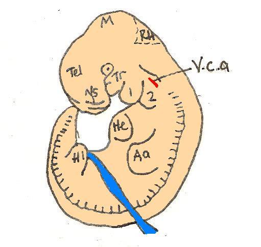 | 511 × 484 (29 KB) | Z3252340 (talk | contribs) | Reverted to version as of 02:05, 20 September 2009 |
| 12:05, 20 September 2009 |  | 511 × 484 (29 KB) | Z3252340 (talk | contribs) | Reverted to version as of 02:05, 20 September 2009 | |
| 12:05, 20 September 2009 |  | 511 × 484 (29 KB) | Z3252340 (talk | contribs) | ||
| 12:05, 20 September 2009 |  | 511 × 484 (29 KB) | Z3252340 (talk | contribs) | ||
| 10:29, 16 September 2009 | 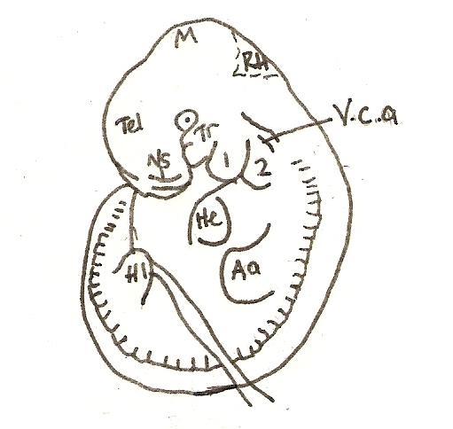 | 511 × 484 (29 KB) | Z3252340 (talk | contribs) | Day 11.5: the lens vesicle is completely separated from the surface. V.ca = vena cardinalis anterior, Tel= telenchephalon, M= mesencephalon, Rh= rhombencephalon, He= heart, Hl= hindlimb bud, Aa= forelimb bud, 1= 1st branchial bar, 2= 2nd branchial bar, T |
You cannot overwrite this file.
File usage
The following 4 pages use this file: