File:Crowder1957 plate03.jpg
Original file (1,280 × 1,416 pixels, file size: 689 KB, MIME type: image/jpeg)
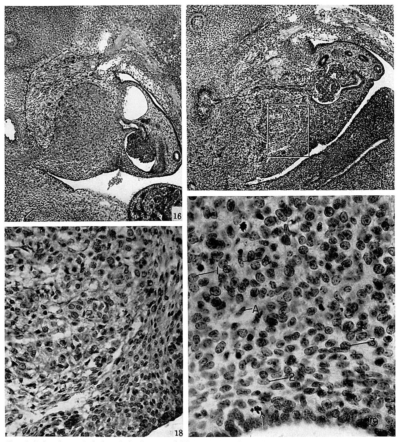
Plate 3
Fig. 16. li.mhr_vn from end of 5th week, Slt{t\\'lI1_$1 ;1 relnztrlt-.thle inc|'e;1.~'4e in the .~;i7.e nl the gluml and p:n'ticul;trl_v in the ntnuunt nf the 5}-'11l}l:1lll(fllC nerve [lHSllt_' (outlined hy hruken line). wliieh lies along the tlursnl and l‘t1L‘Lll;Il sttrfuee of the primnrtlium. tu:m_v filters uml their cells infiltrating it. Sectinn |1L';lI' the eeplmlie pule. Nu. fr,-._;2. 13-2-3. l‘.;u'|_\' lun'i'/.un xvii. mm. H mm.
Fig. 17. Iimln-_m mare :I(l\';lI'lt_‘L'(l than the preceding. Final stage of the lI1\’;ISl(Jll nli nettrztl elements intn the ;ult‘en;1l primnr- tlium. Ct.II‘IlL':ll cells are SCP.'ll‘:lIt.‘(l into isl:tntl.~'. hy the neural tis- sue. except in the \'t.?I1ll‘()l:l[t_'l';ll m;u'gin. 'l‘_vpe (I—ll cells still 11:155- ing from lv§m\'n1:1n'.~e enpstlle into the nitlus urea henenth the tttL'.snItt'}>lt|'iC ;n'ter}' :|t :n‘1‘<m'. Nu. ()f',§:\l. If»;-5. l*luri'/.nn xvii,
Itm. I.2.H mm.
Fig. 18. IH. .-\re;t lI‘|Cllltlt‘.(l within rectangle in figure :7. Two
reginns nf pritnnrtlittm are tlistiltct. :\l)t)\'L' and tu left. urea in- liltrzttetl l‘.}’ neural elements which .\lll'[’Ulll1(l gruups uf t3.'pe.~‘. (ll (ll1(l ('Z—ll| cells. Helnw untl tn right, types Cl and C-Ill cells are heing prnliferzttetl frmn the eelmnie epithelium. ln this clemer area are ;1l.~;n m;tn_\' type C-ll cells. 2-2’ 400.
Fig. 19. Sagittal section «if .'ll'l emhr_vn later in lmri'/..un xvii than the nther.~4 lllllSlI';llL‘(l. taken frmn near the center of luwer pole pf the. gluntl :m(l inelutlin_s: the ventral thirtl. (jelumie epithelium ill lower hurtler. All the cellular elements of the
- ulren
- tl. Cell types (I-l (I) untl C-lll (3) m'igin;ttin;: in the
eelnmie epithelium lurm curds in the e.\p;uuling glztntl. iliypc CH (2) cells lie }uu‘;1llel In the cutter stn'l'uee. nntl :1 few nerte cells. uml lll)L'l‘.\ (.-I") C.\'lL‘I'I(l hetween the eurtienl cells. No. (n~_‘,|:}, tf:-l-3- Hurizctm xvii. _‘f-"'I’mn. tn.=\" mm. Arrcm. lltltn.~»'l.\ in eelnmic epithelium.
- Links: fig 1 | fig 2 | fig 3 | fig 4 | fig 5 | fig 6 | fig 7 | fig 1-7 | plate 1 | plate 2 | plate 3 | 1957 Crowder | Adrenal Development
References
Crowder RE. The development of the adrenal gland in man, with special reference to origin and ultimate location of cell types and evidence in favor of the "cell migration" theory. (1957) Contrib. Embryol., Carnegie Inst. Wash. 36, 193-210.
Cite this page: Hill, M.A. (2024, April 24) Embryology Crowder1957 plate03.jpg. Retrieved from https://embryology.med.unsw.edu.au/embryology/index.php/File:Crowder1957_plate03.jpg
- © Dr Mark Hill 2024, UNSW Embryology ISBN: 978 0 7334 2609 4 - UNSW CRICOS Provider Code No. 00098G
File history
Click on a date/time to view the file as it appeared at that time.
| Date/Time | Thumbnail | Dimensions | User | Comment | |
|---|---|---|---|---|---|
| current | 21:26, 21 June 2017 | | 1,280 × 1,416 (689 KB) | Z8600021 (talk | contribs) | |
| 21:25, 21 June 2017 | 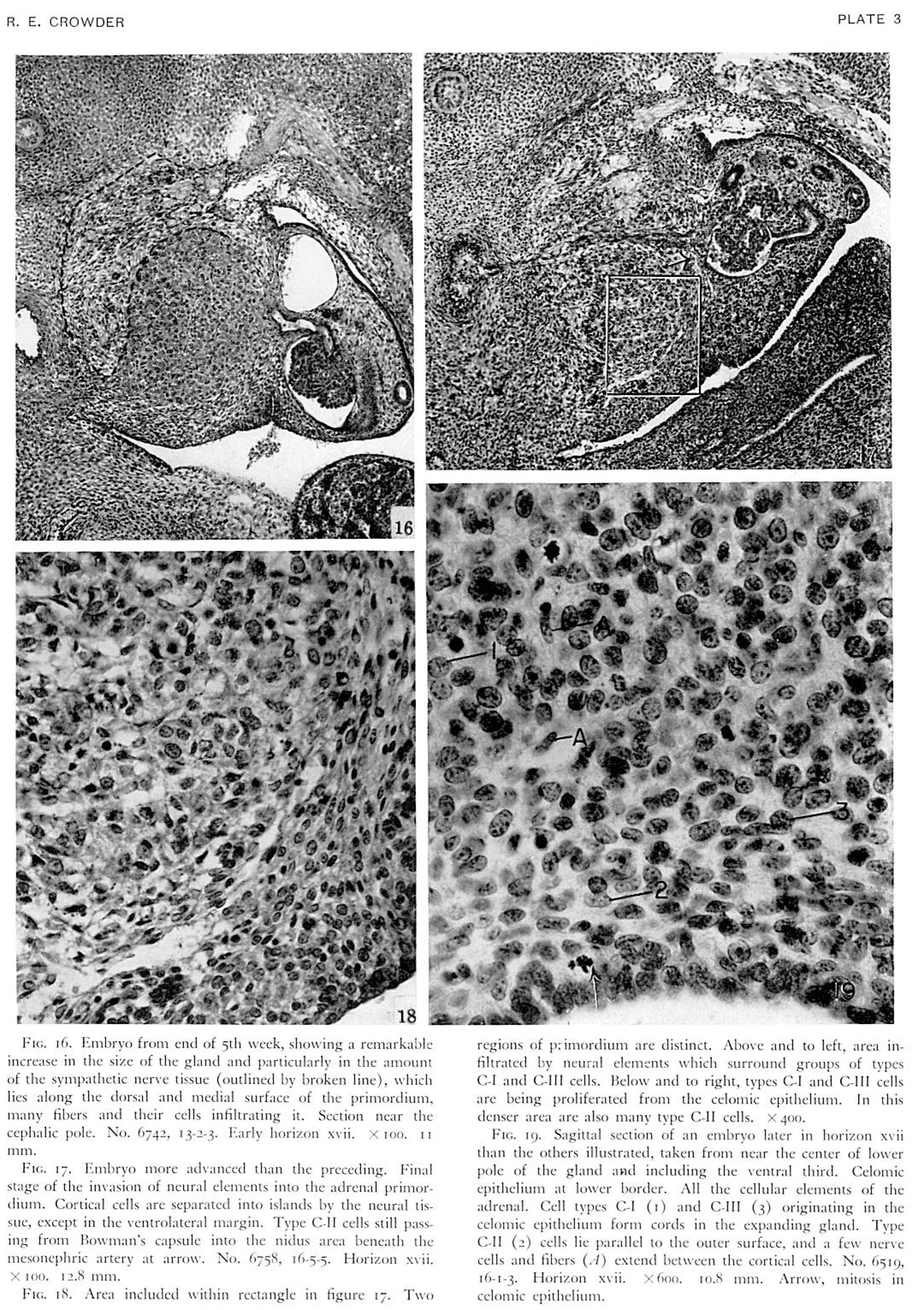 | 2,159 × 3,061 (1.86 MB) | Z8600021 (talk | contribs) | ==Plate 3== {{Crowder1957 figures}} |
You cannot overwrite this file.
File usage
The following page uses this file: