File:Crowder1957 plate03.jpg: Difference between revisions
Original file (1,280 × 1,416 pixels, file size: 689 KB, MIME type: image/jpeg)
mNo edit summary |
m (→Plate 3) |
||
| Line 1: | Line 1: | ||
==Plate 3== | ==Plate 3== | ||
Fig. 16 | Fig. 16 | ||
Fig. 17. | Fig. 17. | ||
Fig. 18 | Fig. 18. | ||
Fig. 19. Sagittal section «if .'ll'l emhr_vn later in lmri'/..un xvii | Fig. 19. Sagittal section «if .'ll'l emhr_vn later in lmri'/..un xvii | ||
Latest revision as of 23:19, 21 June 2017
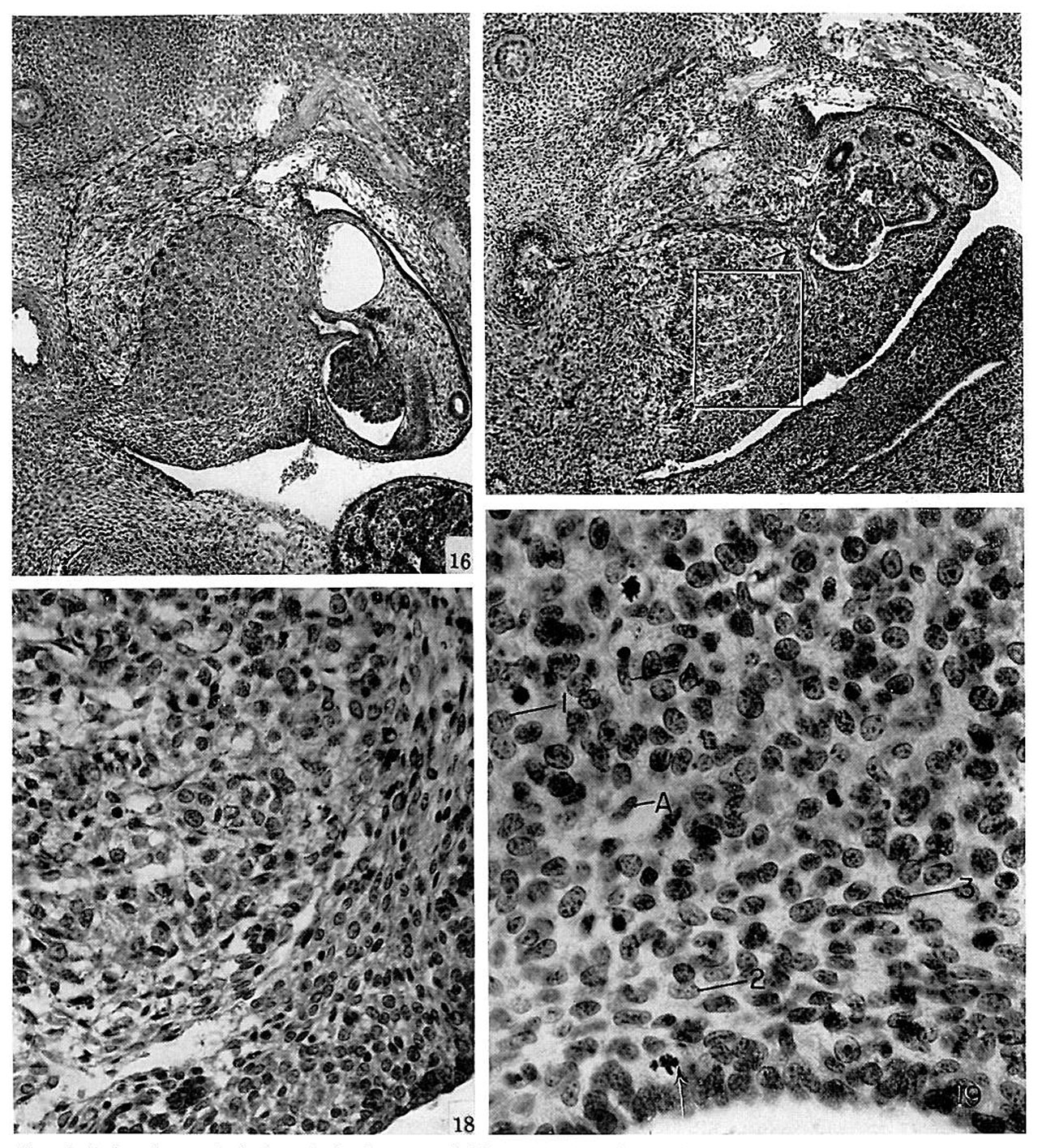
Plate 3
Fig. 16
Fig. 17.
Fig. 18.
Fig. 19. Sagittal section «if .'ll'l emhr_vn later in lmri'/..un xvii than the nther.~4 lllllSlI';llL‘(l. taken frmn near the center of luwer pole pf the. gluntl :m(l inelutlin_s: the ventral thirtl. (jelumie epithelium ill lower hurtler. All the cellular elements of the
- ulren
- tl. Cell types (I-l (I) untl C-lll (3) m'igin;ttin;: in the
eelnmie epithelium lurm curds in the e.\p;uuling glztntl. iliypc CH (2) cells lie }uu‘;1llel In the cutter stn'l'uee. nntl :1 few nerte cells. uml lll)L'l‘.\ (.-I") C.\'lL‘I'I(l hetween the eurtienl cells. No. (n~_‘,|:}, tf:-l-3- Hurizctm xvii. _‘f-"'I’mn. tn.=\" mm. Arrcm. lltltn.~»'l.\ in eelnmic epithelium.
- Links: fig 1 | fig 2 | fig 3 | fig 4 | fig 5 | fig 6 | fig 7 | fig 1-7 | plate 1 | plate 2 | plate 3 | 1957 Crowder | Adrenal Development
References
Crowder RE. The development of the adrenal gland in man, with special reference to origin and ultimate location of cell types and evidence in favor of the "cell migration" theory. (1957) Contrib. Embryol., Carnegie Inst. Wash. 36, 193-210.
Cite this page: Hill, M.A. (2024, April 18) Embryology Crowder1957 plate03.jpg. Retrieved from https://embryology.med.unsw.edu.au/embryology/index.php/File:Crowder1957_plate03.jpg
- © Dr Mark Hill 2024, UNSW Embryology ISBN: 978 0 7334 2609 4 - UNSW CRICOS Provider Code No. 00098G
File history
Click on a date/time to view the file as it appeared at that time.
| Date/Time | Thumbnail | Dimensions | User | Comment | |
|---|---|---|---|---|---|
| current | 21:26, 21 June 2017 | | 1,280 × 1,416 (689 KB) | Z8600021 (talk | contribs) | |
| 21:25, 21 June 2017 | 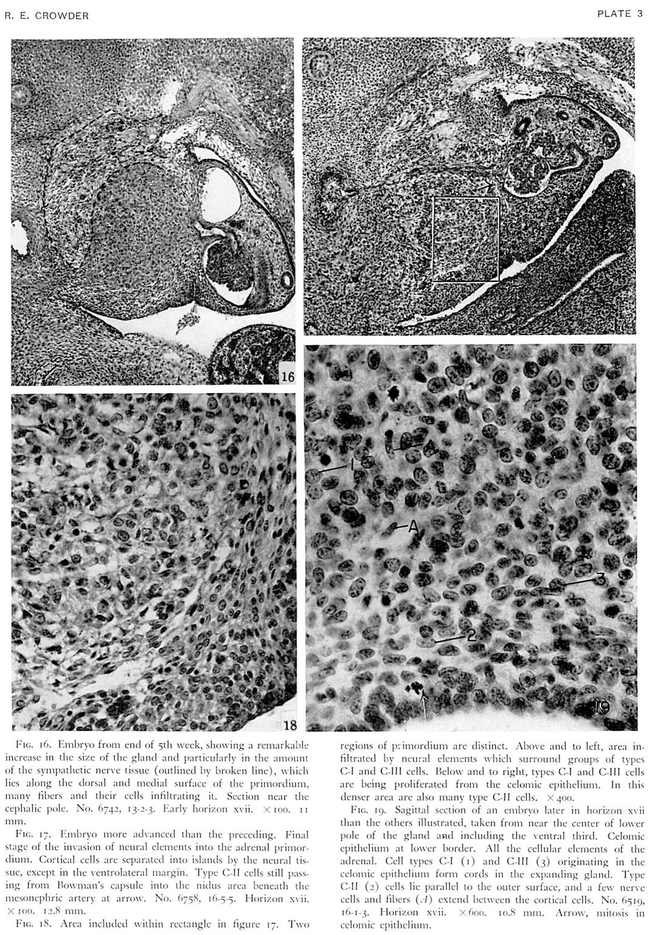 | 2,159 × 3,061 (1.86 MB) | Z8600021 (talk | contribs) | ==Plate 3== {{Crowder1957 figures}} |
You cannot overwrite this file.
File usage
The following page uses this file: