File:Corpus luteum lutein cells.jpg
Corpus_luteum_lutein_cells.jpg (450 × 600 pixels, file size: 104 KB, MIME type: image/jpeg)
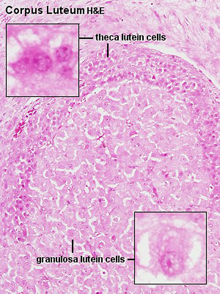
Ovary - Corpus Luteum
Histology image shows the theca lutein cells and granulosa lutein cells. These cells work together in the production of ovarian hormones that support the initial pregnancy.
Theca Lutein Cells
- the darker stained cells.
- derived from the theca interna of the original follicle.
- lack microvilli on the surface.
- lack the aromatase enzyme.
- produce androgens for the granulosa lutein cells to convert.
Granulosa Lutein Cells
- the lighter stained cells.
- derived from the granulosa cells of the original follicle.
- contain aromatase enzyme.
- produce estrogen and progesterone from the androgens produced by the theca lutein cells.
Links: Histology | Histology Stains | Blue Histology images copyright Lutz Slomianka 1998-2009. The literary and artistic works on the original Blue Histology website may be reproduced, adapted, published and distributed for non-commercial purposes. See also the page Histology Stains.
Cite this page: Hill, M.A. (2024, April 24) Embryology Corpus luteum lutein cells.jpg. Retrieved from https://embryology.med.unsw.edu.au/embryology/index.php/File:Corpus_luteum_lutein_cells.jpg
- © Dr Mark Hill 2024, UNSW Embryology ISBN: 978 0 7334 2609 4 - UNSW CRICOS Provider Code No. 00098G
Histology image H&E high power Clu10he.jpg
File history
Click on a date/time to view the file as it appeared at that time.
| Date/Time | Thumbnail | Dimensions | User | Comment | |
|---|---|---|---|---|---|
| current | 16:36, 6 May 2012 | | 450 × 600 (104 KB) | Z8600021 (talk | contribs) | |
| 10:16, 3 August 2009 | 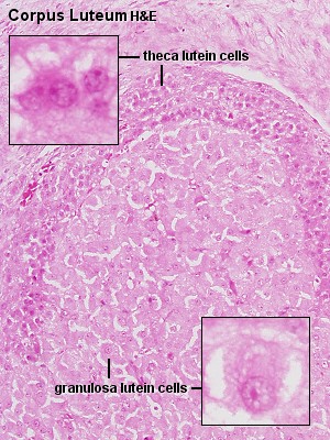 | 300 × 400 (61 KB) | MarkHill (talk | contribs) | Corpus luteum Showing theca lutein cells and granulosa lutein cells Histology image H&E high power Image Source: Lutz Slomianka, UWA Blue Histology Clu10he.jpg http://www.lab.anhb.uwa.edu.au/mb140/CorePages/FemaleRepro/femalerepro.htm#Corpus |
You cannot overwrite this file.