File:Corpus luteum lutein cells.jpg
Corpus_luteum_lutein_cells.jpg (450 × 600 pixels, file size: 104 KB, MIME type: image/jpeg)
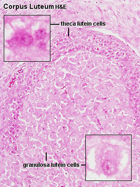
Ovary - Corpus Luteum
Histology image shows the theca lutein cells and granulosa lutein cells. These cells work together in the production of for
Theca Lutein Cells
- the darker stained cells.
- derived from the theca interna of the original follicle.
- lack microvilli on the surface.
- lack the aromatase enzyme.
- produce androgens for the granulosa lutein cells to convert.
Granulosa Lutein Cells
- the lighter stained cells.
- derived from the granulosa cells of the follicle.
- contain aromatase enzyme.
- produce estrogen and progesterone from the androgens produced by the theca lutein cells.
Links: Histology | Histology Stains | Blue Histology images copyright Lutz Slomianka 1998-2009. The literary and artistic works on the original Blue Histology website may be reproduced, adapted, published and distributed for non-commercial purposes. See also the page Histology Stains.
Cite this page: Hill, M.A. (2024, April 16) Embryology Corpus luteum lutein cells.jpg. Retrieved from https://embryology.med.unsw.edu.au/embryology/index.php/File:Corpus_luteum_lutein_cells.jpg
- © Dr Mark Hill 2024, UNSW Embryology ISBN: 978 0 7334 2609 4 - UNSW CRICOS Provider Code No. 00098G
Histology image H&E high power Clu10he.jpg
File history
Click on a date/time to view the file as it appeared at that time.
| Date/Time | Thumbnail | Dimensions | User | Comment | |
|---|---|---|---|---|---|
| current | 16:36, 6 May 2012 | | 450 × 600 (104 KB) | Z8600021 (talk | contribs) | |
| 10:16, 3 August 2009 |  | 300 × 400 (61 KB) | MarkHill (talk | contribs) | Corpus luteum Showing theca lutein cells and granulosa lutein cells Histology image H&E high power Image Source: Lutz Slomianka, UWA Blue Histology Clu10he.jpg http://www.lab.anhb.uwa.edu.au/mb140/CorePages/FemaleRepro/femalerepro.htm#Corpus |
You cannot overwrite this file.