File:Congdon1922-27-28.jpg

Original file (997 × 612 pixels, file size: 68 KB, MIME type: image/jpeg)
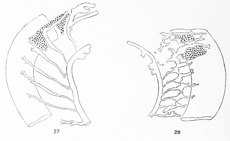
Figs. 27 and 28. Stages in the Formation of the Vertebral Artery
Figure 27
(embryo No. 721, 9 mm.), two segmental arteries arc interrupted but no anastomoses have yet formed between them.
Figure 28
(embryo No. 143, 9 mm.), retrocostal anastomoses have formed between all but the first and second segmental arteries.
- Human Aortic Arch 1922: Table 1 | Fig. 1-16 | Fig 17 | Fig 18-25 | Fig 18 | Fig 19 | Fig 20 | Fig 21 | Fig 22 | Fig 23 | Fig 24 | Fig 25 | Fig 26 | Fig 27-28 | Fig 29 | Fig 30 | Fig 31 | Fig 32 | Fig 33 | Fig 34 | Fig 35 | Fig 36 | Fig 37 | Fig 38 | Fig 39 | Fig 40 | Plate 1 | Plate 2 | Plate 3 | Carnegie No.68 | Volume XIV | Contributions to Embryology | Historic Disclaimer | Cardiovascular Development | Respiratory Development
Reference
Congdon ED. Transformation of the aortic-arch system during the development of the human embryo. (1922) Contrib. Embryol., Carnegie Inst. Wash. Publ 277, 14:47-110.
Cite this page: Hill, M.A. (2024, April 20) Embryology Congdon1922-27-28.jpg. Retrieved from https://embryology.med.unsw.edu.au/embryology/index.php/File:Congdon1922-27-28.jpg
- © Dr Mark Hill 2024, UNSW Embryology ISBN: 978 0 7334 2609 4 - UNSW CRICOS Provider Code No. 00098G
| Historic Disclaimer - information about historic embryology pages |
|---|
 |
File history
Click on a date/time to view the file as it appeared at that time.
| Date/Time | Thumbnail | Dimensions | User | Comment | |
|---|---|---|---|---|---|
| current | 17:59, 7 May 2011 | | 997 × 612 (68 KB) | S8600021 (talk | contribs) | ==Figs. 27 and 28. Stages in the formation of the vertebral artery== In figure 27 (embryo No. 721, 9 mm.), two segmental arteries arc interrupted but no anastomoses have yet formed between them. In figure 28 (embryo No. 143, 9 mm.), retrocostal anastom |
You cannot overwrite this file.
File usage
The following page uses this file: