File:Cochlea MRI 02.jpg: Difference between revisions
No edit summary |
No edit summary |
||
| Line 7: | Line 7: | ||
Adult cochlear length varied between 17 and 26.5 millimeters. | Adult cochlear length varied between 17 and 26.5 millimeters. | ||
:'''Links:''' [[:File:Cochlea MRI 01.jpg|Cochlea MRI 1]] | [[:File:Cochlea_MRI_02.jpg|Cochlea MRI 2]] | [[Hearing_-_Inner_Ear_Development|Inner Ear Development]] | [[Sensory_-_Hearing_and_Balance_Development|Hearing and Balance Development]] | [[Magnetic Resonance Imaging]] | |||
===Reference=== | |||
<pubmed>19575114</pubmed>| [http://www.scielo.br/scielo.php?script=sci_arttext&pid=S0034-72992009000200017&lng=en&nrm=iso&tlng=en Braz J Otorhinolaryngol.] | <pubmed>19575114</pubmed>| [http://www.scielo.br/scielo.php?script=sci_arttext&pid=S0034-72992009000200017&lng=en&nrm=iso&tlng=en Braz J Otorhinolaryngol.] | ||
All the content of the journal, except where otherwise noted, is licensed under a [http://creativecommons.org/licenses/by-nc/3.0/ Creative Commons License] | All the content of the journal, except where otherwise noted, is licensed under a [http://creativecommons.org/licenses/by-nc/3.0/ Creative Commons License] | ||
Original File name: Fig 6. http://www.scielo.br/img/revistas/rboto/v75n2/en_a17fig06.gif | |||
[[Category:Hearing]] [[Category:Magnetic Resonance Imaging]] | [[Category:Hearing]] [[Category:Magnetic Resonance Imaging]] | ||
Latest revision as of 04:38, 19 August 2012
Adult Human Cochlea MRI
The 3D reconstructed technique was used to acquire coronal and axial images of the adult inner ear. The coronal section reconstruction was chosen since it increases visibility of the turns of the cochlea.
The investigators used a method to obtain magnetic resonance imaging (MRI) to measure cochlear length from the temporal bones of 6 cadavers. By overlapping digitalized rulers on these images it was possible to measure cochlear length.
Adult cochlear length varied between 17 and 26.5 millimeters.
- Links: Cochlea MRI 1 | Cochlea MRI 2 | Inner Ear Development | Hearing and Balance Development | Magnetic Resonance Imaging
Reference
<pubmed>19575114</pubmed>| Braz J Otorhinolaryngol.
All the content of the journal, except where otherwise noted, is licensed under a Creative Commons License
Original File name: Fig 6. http://www.scielo.br/img/revistas/rboto/v75n2/en_a17fig06.gif
File history
Click on a date/time to view the file as it appeared at that time.
| Date/Time | Thumbnail | Dimensions | User | Comment | |
|---|---|---|---|---|---|
| current | 12:22, 11 August 2010 | 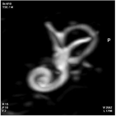 | 381 × 380 (12 KB) | S8600021 (talk | contribs) | ==Adult Human Cochlea MRI== The 3D reconstructed technique was used to acquire coronal and axial images of the adult inner ear. The coronal section reconstruction was chosen since it increases visibility of the turns of the cochlea. The investigators us |
You cannot overwrite this file.
File usage
The following 2 pages use this file: