File:Cat inner ear MicroCT.jpg: Difference between revisions
(MicroCT imaging of an unstained cat inner ear for visualization of mineralized and soft tissues. Voxel size was 10 μm isotropic. (E) Volume rendering from the soft tissue volume, revealing nerves, bone marrow, and the membranous labyrinth. Legend: am...) |
mNo edit summary |
||
| (One intermediate revision by the same user not shown) | |||
| Line 1: | Line 1: | ||
==Cat inner ear MicroCT== | |||
( | Cat inner ear MicroCT (unstained) for visualization of mineralized and soft tissues. Volume rendering from the soft tissue volume, revealing nerves, bone marrow, and the membranous labyrinth. Voxel size was 10 μm isotropic. | ||
Legend: am, ampulla; bm, bone marrow; co, cochlea; he, helicotrema; nc, cochlear nerve; nf, facial nerve; nv, vestibular nerve; sc, semi-circular canal. | |||
:Links: {{inner ear}} | {{cat}} | |||
===Reference=== | ===Reference=== | ||
{{#pmid:30108474}} | {{#pmid:30108474}} | ||
| Line 12: | Line 16: | ||
{{Footer}} | {{Footer}} | ||
[[Category:Cat]][[Category:Inner Ear]][[Category:Computed Tomography]] | [[Category:Cat]][[Category:Inner Ear]][[Category:Hearing]][[Category:Computed Tomography]] | ||
Latest revision as of 12:52, 6 September 2018
Cat inner ear MicroCT
Cat inner ear MicroCT (unstained) for visualization of mineralized and soft tissues. Volume rendering from the soft tissue volume, revealing nerves, bone marrow, and the membranous labyrinth. Voxel size was 10 μm isotropic.
Legend: am, ampulla; bm, bone marrow; co, cochlea; he, helicotrema; nc, cochlear nerve; nf, facial nerve; nv, vestibular nerve; sc, semi-circular canal.
Reference
Glueckert R, Johnson Chacko L, Schmidbauer D, Potrusil T, Pechriggl EJ, Hoermann R, Brenner E, Reka A, Schrott-Fischer A & Handschuh S. (2018). Visualization of the Membranous Labyrinth and Nerve Fiber Pathways in Human and Animal Inner Ears Using MicroCT Imaging. Front Neurosci , 12, 501. PMID: 30108474 DOI.
Copyright
© 2018 Glueckert, Johnson Chacko, Schmidbauer, Potrusil, Pechriggl, Hoermann, Brenner, Reka, Schrott-Fischer and Handschuh. This is an open-access article distributed under the terms of the Creative Commons Attribution License (CC BY). The use, distribution or reproduction in other forums is permitted, provided the original author(s) and the copyright owner(s) are credited and that the original publication in this journal is cited, in accordance with accepted academic practice. No use, distribution or reproduction is permitted which does not comply with these terms.
FIGURE 4. g004.jpg
Cite this page: Hill, M.A. (2024, April 25) Embryology Cat inner ear MicroCT.jpg. Retrieved from https://embryology.med.unsw.edu.au/embryology/index.php/File:Cat_inner_ear_MicroCT.jpg
- © Dr Mark Hill 2024, UNSW Embryology ISBN: 978 0 7334 2609 4 - UNSW CRICOS Provider Code No. 00098G
File history
Click on a date/time to view the file as it appeared at that time.
| Date/Time | Thumbnail | Dimensions | User | Comment | |
|---|---|---|---|---|---|
| current | 12:24, 6 September 2018 | 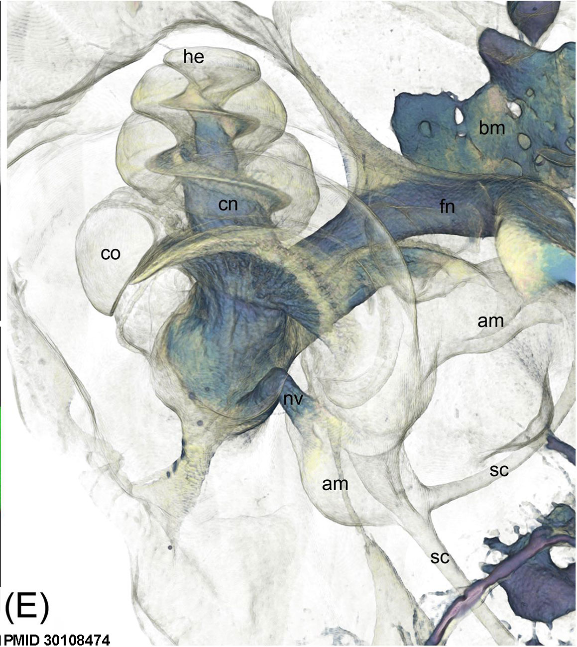 | 1,159 × 1,300 (266 KB) | Z8600021 (talk | contribs) | MicroCT imaging of an unstained cat inner ear for visualization of mineralized and soft tissues. Voxel size was 10 μm isotropic. (E) Volume rendering from the soft tissue volume, revealing nerves, bone marrow, and the membranous labyrinth. Legend: am... |
You cannot overwrite this file.
File usage
The following page uses this file: