File:CSt3.jpg: Difference between revisions
From Embryology
No edit summary |
|||
| (14 intermediate revisions by one other user not shown) | |||
| Line 1: | Line 1: | ||
==Human Embryo Carnegie stage 3== | |||
Human Blastocyst "hatching" from zona pellucida, in early [[Embryonic Development]] designated as [[Carnegie stage 3]]. | Human Blastocyst "hatching" from zona pellucida, in early [[Embryonic Development]] designated as [[Carnegie stage 3]]. | ||
''' | The '''zona pellucida''' is shown to the left of the image and the '''blastocyst''' is to the right of image. | ||
Note: | '''Note:''' | ||
* the small opening in the zona pellucida through which the blastocyst is hatching | * the small opening in the zona pellucida through which the blastocyst is hatching | ||
* the flattened trophoblast cells forming the outer cell layer of the blastocyst | * the flattened trophoblast cells forming the outer cell layer of the blastocyst | ||
| Line 9: | Line 11: | ||
* the blastocoel forming a large fluid-filled space within the blastocyst | * the blastocoel forming a large fluid-filled space within the blastocyst | ||
:'''Links:''' [[:File:CSt3.jpg|unlabeled image]] | [[:File:Human carnegie stage 3 label.jpg|labeled image]] | [[Carnegie stage 3]] | [[Blastocyst Development]] | [[Week 1]] | |||
===Reference=== | |||
<pubmed>16929302</pubmed>| [http://www.nature.com/nature/journal/v444/n7118/full/nature05142.html Nature] | |||
====Copyright==== | |||
Adapted by permission from Macmillan Publishers Ltd (copyright 2006) | Adapted by permission from Macmillan Publishers Ltd (copyright 2006) | ||
[[Category:Human Embryo]] [[Category:Carnegie Stage]] [[Category:Carnegie Stage 3]] [[Category:Week 1]] | Original image name: Figure 1 part b, Development of a blastomere-biopsied embryo into a hatching blastocyst. http://www.nature.com/nature/journal/v444/n7118/fig_tab/nature05142_F1.html (image extracted from full figure and adjusted in size and contrast) | ||
[[Category:Human Embryo]] [[Category:Carnegie Stage]] [[Category:Carnegie Stage 3]] [[Category:Week 1]] [[Category:Blastocyst]] | |||
Latest revision as of 13:38, 29 November 2012
Human Embryo Carnegie stage 3
Human Blastocyst "hatching" from zona pellucida, in early Embryonic Development designated as Carnegie stage 3.
The zona pellucida is shown to the left of the image and the blastocyst is to the right of image.
Note:
- the small opening in the zona pellucida through which the blastocyst is hatching
- the flattened trophoblast cells forming the outer cell layer of the blastocyst
- the inner cell mass shown in the centre of the image and on the left-hand wall of the blastocyst
- the blastocoel forming a large fluid-filled space within the blastocyst
- Links: unlabeled image | labeled image | Carnegie stage 3 | Blastocyst Development | Week 1
Reference
<pubmed>16929302</pubmed>| Nature
Copyright
Adapted by permission from Macmillan Publishers Ltd (copyright 2006)
Original image name: Figure 1 part b, Development of a blastomere-biopsied embryo into a hatching blastocyst. http://www.nature.com/nature/journal/v444/n7118/fig_tab/nature05142_F1.html (image extracted from full figure and adjusted in size and contrast)
File history
Click on a date/time to view the file as it appeared at that time.
| Date/Time | Thumbnail | Dimensions | User | Comment | |
|---|---|---|---|---|---|
| current | 13:07, 9 May 2011 | 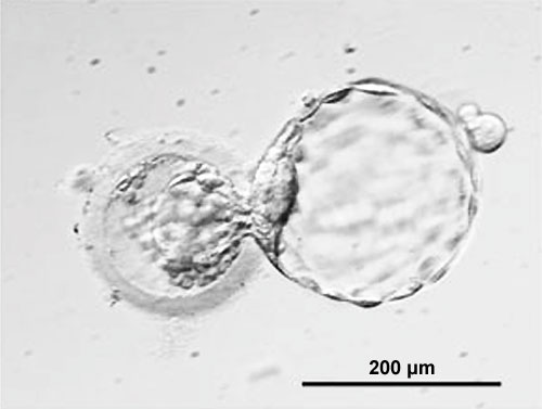 | 500 × 377 (20 KB) | S8600021 (talk | contribs) | |
| 12:55, 9 May 2011 | 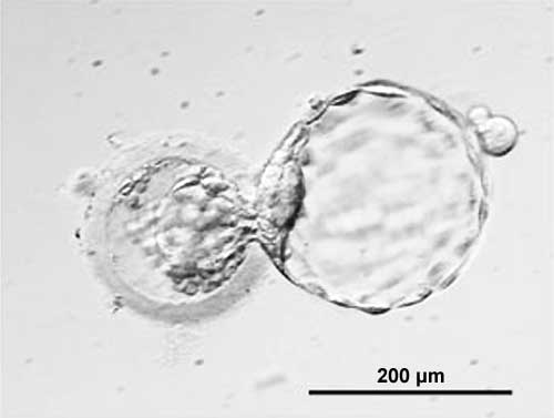 | 500 × 377 (12 KB) | S8600021 (talk | contribs) | ||
| 12:54, 9 May 2011 | 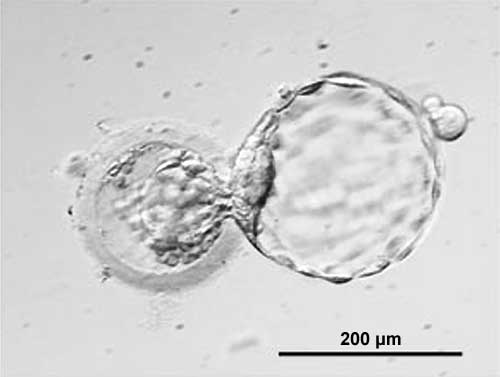 | 500 × 377 (12 KB) | S8600021 (talk | contribs) | ||
| 12:32, 2 August 2009 | 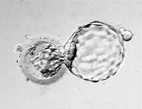 | 285 × 219 (11 KB) | S8600021 (talk | contribs) | Image source: Klimanskaya I, Chung Y, Becker S, Lu SJ, Lanza R. Human embryonic stem cell lines derived from single blastomeres. Nature. 2006 Aug 23 [http://www.ncbi.nlm.nih.gov:80/entrez/query.fcgi?cmd=Retrieve&db=pubmed&dopt=Abstract&list_uids=16929302 |
You cannot overwrite this file.
File usage
The following 49 pages use this file:
- 2009 Lab 10
- 2009 Lecture 3
- 2010 BGD Lecture - Development of the Embryo/Fetus 2
- 2010 BGD Practical 3 - Early Cell Division
- 2010 BGD Practical 3 - Week 1 Summary
- 2010 BGD Practical 3 - Week 3 Summary
- 2010 BGD Practical 6 - Week 8
- 2010 BGD Tutorial - Applied Embryology and Teratology
- 2010 Lab 2
- 2010 Lecture 3
- 2010 Lecture 6
- 2011 Group Project 11
- ANAT2341 Lab 2 - Week 1
- Abnormal Development - Environmental
- Abnormal Development - Illegal Drugs
- Abnormal Development - Teratogens
- Abnormal Development - Twinning
- BGDA Lecture - Development of the Embryo/Fetus 2
- BGDA Practical 3 - Early Cell Division
- BGDA Practical 3 - Week 1 Summary
- BGDA Practical 3 - Week 2 Summary
- BGDA Practical 3 - Week 3 Summary
- BGDA Practical 7 - Week 8
- BGD Tutorial - Applied Embryology and Teratology
- Carnegie stage 3
- Embryonic Development
- Foundations Practical - Critical Periods
- Human Abnormal Development
- K12 Stem Cells
- Lecture - Ectoderm Development
- Lecture - Fetal Development
- Lecture - Week 1 and 2 Development
- Placenta - Membranes
- Stem Cells
- Timeline - Human Embryonic Stem Cell Research
- Timeline human development
- Week 1
- Week 1 - Abnormalities
- Week 2
- Zona pellucida
- Talk:Timeline human development
- File:Human-critical periods of development.jpg
- File talk:Human-critical periods.jpg
- Template:Critical Periods table
- Template:First Trimester Timeline
- Template:First Trimester Timeline collapsable table
- Template:Monoygotic Twinning Table
- Template:Week 1 table
- Template talk:First Trimester Timeline