File:CNS primary vesicles.jpg
Original file (987 × 562 pixels, file size: 49 KB, MIME type: image/jpeg)
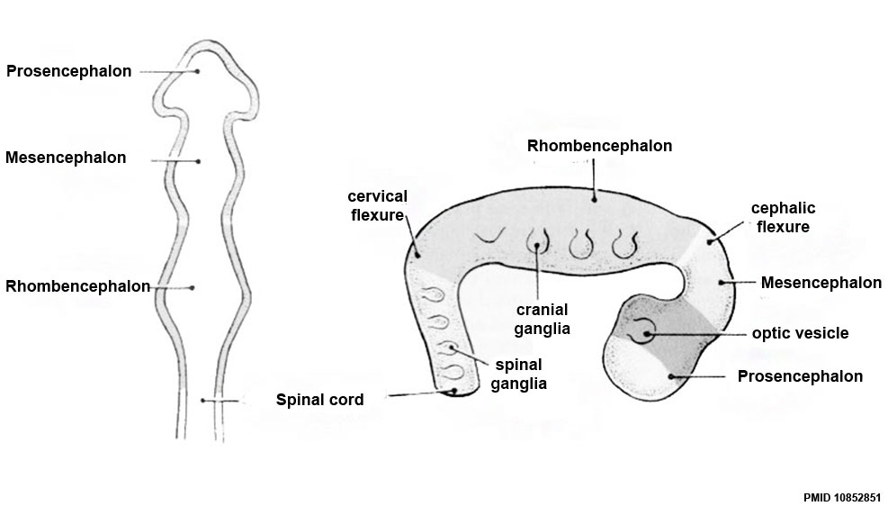
Central Nervous System - primary vesicles
These 3 primary brain vesicles develop initially due to the neural plate being broader at the cranial (brain) end that the narrower caudal (spinal cord) end. When the plate fuses to form a tube, these 3 initial expansions (vesicles) result.
The development of the three primary brain vesicles on GD 10.5 in rats and GD 26 ± 1 in humans. The corresponding shading between panels illustrates the earlier origins of different regions from the three original brain vesicles with a horizontal and lateral view, respectively. Later they will develop into the 5 secondary vesicles.
Three primary brain vesicles: forebrain ( prosencephalon) - midbrain (mesencephalon) - hindbrain (rhombencephalon)
Original file name: Figure-3AB-PMID10852851.jpg
Reference
<pubmed>10852851</pubmed>| Environmental Health Perspectives | PMC: 1637807
File history
Click on a date/time to view the file as it appeared at that time.
| Date/Time | Thumbnail | Dimensions | User | Comment | |
|---|---|---|---|---|---|
| current | 16:48, 18 May 2017 | | 987 × 562 (49 KB) | Z8600021 (talk | contribs) | |
| 16:18, 5 April 2017 | 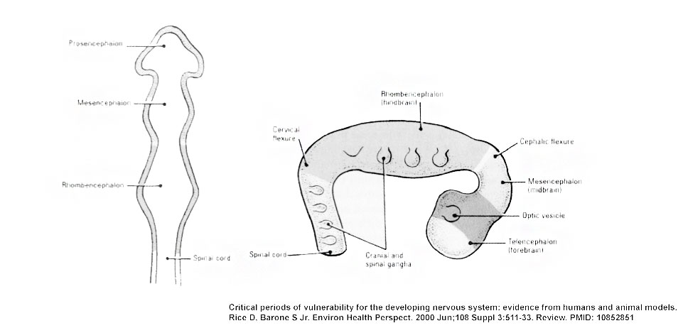 | 961 × 462 (45 KB) | Z8600021 (talk | contribs) | ||
| 01:01, 11 August 2009 | 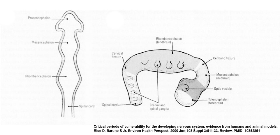 | 961 × 462 (50 KB) | MarkHill (talk | contribs) | CNS primary vesicles cartoon Figure-3AB-PMID10852851.jpg Image Source: Critical periods of vulnerability for the developing nervous system: evidence from humans and animal models. Rice D, Barone S Jr. Environ Health Perspect. 2000 Jun;108 Suppl 3:511-33 |
You cannot overwrite this file.
File usage
The following 36 pages use this file:
- 2009 Lecture 6
- 2010 BGD Lecture - Development of the Embryo/Fetus 2
- 2010 BGD Practical 6 - Week 4
- 2010 Lab 3
- 2010 Lecture 6
- 2011 Lab 3 - Week 4
- ANAT2341 Lab 3 - Week 4
- BGDA Lecture - Development of the Embryo/Fetus 2
- BGDA Lecture - Development of the Nervous System
- BGDA Practical 7 - Week 4
- Brain Awareness Week 2012
- K12 Brain Awareness Week
- Lecture - Ectoderm Development
- Lecture - Neural Development
- Neural - Amygdala Development
- Neural - Basal Ganglia Development
- Neural - Cerebellum Development
- Neural - Cerebrum Development
- Neural - Diencephalon Development
- Neural - Hippocampus Development
- Neural - Medulla Oblongata Development
- Neural - Mesencephalon Development
- Neural - Metencephalon Development
- Neural - Myelencephalon Development
- Neural - Pons Development
- Neural - Prosencephalon Development
- Neural - Rhinencephalon Development
- Neural - Rhombencephalon Development
- Neural - Spinal Cord Development
- Neural - Tectum Development
- Neural - Telencephalon Development
- Neural - Thalamus Development
- Neural System Development
- P
- Talk:2011 Lab 3
- Talk:BGDA Lecture - Development of the Nervous System