File:Brown012.jpg: Difference between revisions
From Embryology
({{Template:Brown 1906 Figures}} Category:Pig) |
No edit summary |
||
| Line 1: | Line 1: | ||
==Fig. 12. Horizontal section through eye of a pig== | |||
Magnified 730 times. | |||
{{Template:Brown 1906 Figures}} | {{Template:Brown 1906 Figures}} | ||
[[Category:Pig]] | [[Category:Pig]] | ||
Revision as of 21:28, 12 November 2012
Fig. 12. Horizontal section through eye of a pig
Magnified 730 times.
| Historic Disclaimer - information about historic embryology pages |
|---|
 |
Reference
Brown EJ. The embryology anatomy and histology of the eye. (1906) Chicago: Hazlitt & Walker.
Cite this page: Hill, M.A. (2024, April 18) Embryology Brown012.jpg. Retrieved from https://embryology.med.unsw.edu.au/embryology/index.php/File:Brown012.jpg
- © Dr Mark Hill 2024, UNSW Embryology ISBN: 978 0 7334 2609 4 - UNSW CRICOS Provider Code No. 00098G
File history
Click on a date/time to view the file as it appeared at that time.
| Date/Time | Thumbnail | Dimensions | User | Comment | |
|---|---|---|---|---|---|
| current | 15:10, 14 February 2011 | 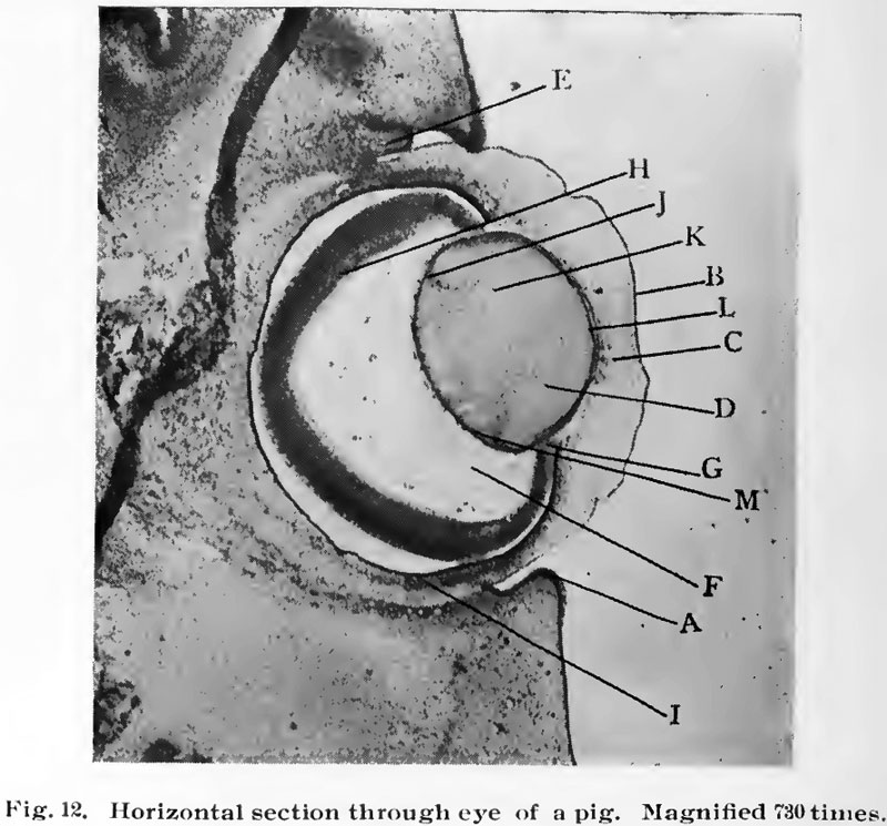 | 800 × 745 (105 KB) | S8600021 (talk | contribs) | {{Template:Brown 1906 Figures}} Category:Pig |
You cannot overwrite this file.
File usage
The following 2 pages use this file: