File:Brown003.jpg: Difference between revisions
({{Template:Brown 1906 Figures}}) |
No edit summary |
||
| (One intermediate revision by one other user not shown) | |||
| Line 1: | Line 1: | ||
==Fig. 3. Vertical Section through Head of Foetal Pig, 2 mm long== | |||
Magnified 2,500 times. | |||
As this stalk grows outward, the anterior portion rises upward, as shown in vertical section at A, Fig. 3. When the optic stalk comes near to the surface, the anterior portion enlarges, as shown at C, Fig. i. Also when the optic stalk encroaches on the surface, it stimulates the epithelial cells forming- the skin and they multiply rapidly (see B, Figs. I and 3), and the anterior wall of the primary optic vesicle invaginates and passes inside of the vesicle. (See A, Fig. 2, and C, Fig. 3.) This invagination might be likened to the denting of a hollow rubber ball. This invaginated portion forms the secondary optic vesicle and it is from this that the nine innermost layers of the retina are eventually formed, while the primary optic vesicle only forms the outer or pigment layer. | |||
{{Template:Brown 1906 Figures}} | {{Template:Brown 1906 Figures}} | ||
[[Category:Pig]] | |||
Latest revision as of 07:08, 31 August 2011
Fig. 3. Vertical Section through Head of Foetal Pig, 2 mm long
Magnified 2,500 times.
As this stalk grows outward, the anterior portion rises upward, as shown in vertical section at A, Fig. 3. When the optic stalk comes near to the surface, the anterior portion enlarges, as shown at C, Fig. i. Also when the optic stalk encroaches on the surface, it stimulates the epithelial cells forming- the skin and they multiply rapidly (see B, Figs. I and 3), and the anterior wall of the primary optic vesicle invaginates and passes inside of the vesicle. (See A, Fig. 2, and C, Fig. 3.) This invagination might be likened to the denting of a hollow rubber ball. This invaginated portion forms the secondary optic vesicle and it is from this that the nine innermost layers of the retina are eventually formed, while the primary optic vesicle only forms the outer or pigment layer.
| Historic Disclaimer - information about historic embryology pages |
|---|
 |
Reference
Brown EJ. The embryology anatomy and histology of the eye. (1906) Chicago: Hazlitt & Walker.
Cite this page: Hill, M.A. (2024, April 19) Embryology Brown003.jpg. Retrieved from https://embryology.med.unsw.edu.au/embryology/index.php/File:Brown003.jpg
- © Dr Mark Hill 2024, UNSW Embryology ISBN: 978 0 7334 2609 4 - UNSW CRICOS Provider Code No. 00098G
File history
Click on a date/time to view the file as it appeared at that time.
| Date/Time | Thumbnail | Dimensions | User | Comment | |
|---|---|---|---|---|---|
| current | 13:12, 14 February 2011 | 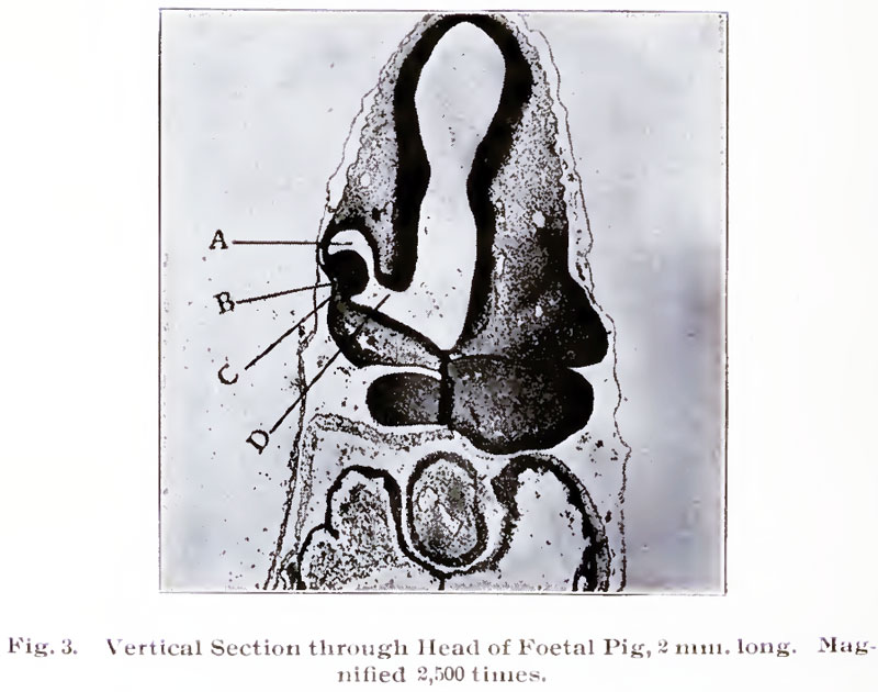 | 800 × 630 (85 KB) | MarkHill (talk | contribs) | {{Template:Brown 1906 Figures}} |
You cannot overwrite this file.