File:Braune 1877 plate 2 fig6.jpg: Difference between revisions
No edit summary |
No edit summary |
||
| Line 16: | Line 16: | ||
{{Braune 1877 | {{Braune 1877 header}} | ||
Latest revision as of 09:14, 10 November 2012
Plate 2 Sagittal Female Fig. 6. Female, set. 35, after childbirth ; ante-flexion
Legendre, xix. 1. Uterus. 2. Bladder. 3. Rectum. 4. Symphysis.
The woman (fig. 6) died immediately after childbirth ; the anteflexion also was recent, brought on by the weight of the heavy body of the uterus, which had a capacity of about two fluid ounces.
The flexion is so considerable that the body and neck of the uterus are almost at a right angle with each other, and on the anterior wall a distinct fold is formed. The posterior wall of the uterus rests on the rectum, and presses on its lumen, &c. The vagina is distended and measures 3.6 inches. The distance of the peritoneum on the posterior wall of the uterus from the perineum is 3.8 inches. These figures consequently considerably exceed those in the preceding case.
The position of the fundus with regard to the firmly-compressed bladder is to be remarked here, so also the position of that portion of the peritoneum which lies between the uterus and bladder on the anterior wall of the vagina. In the normal condition of the uterus its end lies nearest the peritoneum, whereas it is here in the middle. It is further to be noticed that the strong attachment of the posterior portion of the base of the bladder with the neck of the uterus (which as already mentioned is admitted by Courty) is not present in this preparation, otherwise the bladder and urethra lying close down on the uterus would be dragged upwards. Nevertheless, the lax cellular tissue and fascia between these viscera is not so capable of distension that variations in the position of the uterus could continue without any influence on the bladder. We notice here that a large piece of the posterior portion of the base of the bladder has been drawn upwards, a condition which would interfere with the action of the vesical sphincter, and consequently cause an incontinence of urine. The conjugate diameter is very large, 5 inches, and exceeds that in PI. II.
- Sagittal Female: Plate 2A | Plate 2B | Plate 2 Fig.1 | Plate 2 Fig.2 | Plate 2 Fig.3 | Plate 2 Fig.4 | Plate 2 Fig.5 | Plate 2 Fig.6 | Plate 2 Fig.7 | Topographical Anatomy (1877)
| Embryology - 23 Apr 2024 |
|---|
| Google Translate - select your language from the list shown below (this will open a new external page) |
|
العربية | català | 中文 | 中國傳統的 | français | Deutsche | עִברִית | हिंदी | bahasa Indonesia | italiano | 日本語 | 한국어 | မြန်မာ | Pilipino | Polskie | português | ਪੰਜਾਬੀ ਦੇ | Română | русский | Español | Swahili | Svensk | ไทย | Türkçe | اردو | ייִדיש | Tiếng Việt These external translations are automated and may not be accurate. (More? About Translations) |
Braune W. An atlas of topographical anatomy after plane sections of frozen bodies. (1877) Trans. by Edward Bellamy. Philadelphia: Lindsay and Blakiston.
- Plates: 1. Male - Sagittal body | 2. Female - Sagittal body | 3. Obliquely transverse head | 4. Transverse internal ear | 5. Transverse head | 6. Transverse neck | 7. Transverse neck and shoulders | 8. Transverse level first dorsal vertebra | 9. Transverse thorax level of third dorsal vertebra | 10. Transverse level aortic arch and fourth dorsal vertebra | 11. Transverse level of the bulbus aortae and sixth dorsal vertebra | 12. Transverse level of mitral valve and eighth dorsal vertebra | 13. Transverse level of heart apex and ninth dorsal vertebra | 14. Transverse liver stomach spleen at level of eleventh dorsal vertebra | 15. Transverse pancreas and kidneys at level of L1 vertebra | 16. Transverse through transverse colon at level of intervertebral space between L3 L4 vertebra | 17. Transverse pelvis at level of head of thigh bone | 18. Transverse male pelvis | 19. knee and right foot | 20. Transverse thigh | 21. Transverse left thigh | 22. Transverse lower left thigh and knee | 23. Transverse upper and middle left leg | 24. Transverse lower left leg | 25. Male - Frontal thorax | 26. Elbow-joint hand and third finger | 27. Transverse left arm | 28. Transverse left fore-arm | 29. Sagittal female pregnancy | 30. Sagittal female pregnancy | 31. Sagittal female at term
| Historic Disclaimer - information about historic embryology pages |
|---|
 |
File history
Click on a date/time to view the file as it appeared at that time.
| Date/Time | Thumbnail | Dimensions | User | Comment | |
|---|---|---|---|---|---|
| current | 14:17, 31 October 2012 | 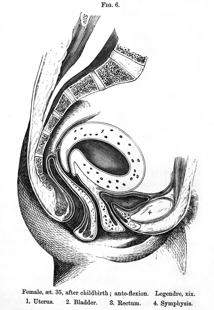 | 690 × 1,000 (144 KB) | Z8600021 (talk | contribs) | {{Braune 1877 footer}} |
You cannot overwrite this file.
File usage
The following 2 pages use this file: