File:Braune 1877 plate 2 fig3.jpg: Difference between revisions
({{Braune 1877 header}} Braune 1877 plate 2 fig3.jpg) |
No edit summary |
||
| Line 1: | Line 1: | ||
{{Braune 1877 | ==Plate 2 Sagittal Female Fig. 3. Female pelvis, set. 35 ; normal ; bladder empty, rectum distended== | ||
Pirogoff, iii, A, 21, fig. 3. 1. Uterus. 2. Bladder. 3. Rectum. 4. Symphysis. | |||
The extent to which the position of the uterus varies with distension and evacuation of the bladder is well shown in figs. 2 and 3. The section in fig. 3 has not gone exactly through the mesial plane, and thus, although the uterus is bisected, the anus and the urethra have escaped division. In fig. 2 we have exactly the opposite conditions, namely, an empty bladder and distended rectum ; consequently the relations of the uterus and vagina are altered. Whereas in fig. 2 the uterus follows the axis of the vagina, in this instance it forms an obtuse angle with it, without, however, being anteverted. No coils of intestine lie between the uterus and rectum. The conjugate diameter is 4.41 inches. | |||
{{Braune 1877 Plate 2}} | |||
{{Braune 1877 footer}} | |||
Braune 1877 plate 2 fig3.jpg | Braune 1877 plate 2 fig3.jpg | ||
Revision as of 08:55, 10 November 2012
Plate 2 Sagittal Female Fig. 3. Female pelvis, set. 35 ; normal ; bladder empty, rectum distended
Pirogoff, iii, A, 21, fig. 3. 1. Uterus. 2. Bladder. 3. Rectum. 4. Symphysis.
The extent to which the position of the uterus varies with distension and evacuation of the bladder is well shown in figs. 2 and 3. The section in fig. 3 has not gone exactly through the mesial plane, and thus, although the uterus is bisected, the anus and the urethra have escaped division. In fig. 2 we have exactly the opposite conditions, namely, an empty bladder and distended rectum ; consequently the relations of the uterus and vagina are altered. Whereas in fig. 2 the uterus follows the axis of the vagina, in this instance it forms an obtuse angle with it, without, however, being anteverted. No coils of intestine lie between the uterus and rectum. The conjugate diameter is 4.41 inches.
- Sagittal Female: Plate 2A | Plate 2B | Plate 2 Fig.1 | Plate 2 Fig.2 | Plate 2 Fig.3 | Plate 2 Fig.4 | Plate 2 Fig.5 | Plate 2 Fig.6 | Plate 2 Fig.7 | Topographical Anatomy (1877)
| Historic Disclaimer - information about historic embryology pages |
|---|
 |
- Braune Plates (1877): 1. Male - Sagittal body | 2. Female - Sagittal body | 3. Obliquely transverse head | 4. Transverse internal ear | 5. Transverse head | 6. Transverse neck | 7. Transverse neck and shoulders | 8. Transverse level first dorsal vertebra | 9. Transverse thorax level of third dorsal vertebra | 10. Transverse level aortic arch and fourth dorsal vertebra | 11. Transverse level of the bulbus aortae and sixth dorsal vertebra | 12. Transverse level of mitral valve and eighth dorsal vertebra | 13. Transverse level of heart apex and ninth dorsal vertebra | 14. Transverse liver stomach spleen at level of eleventh dorsal vertebra | 15. Transverse pancreas and kidneys at level of L1 vertebra | 16. Transverse through transverse colon at level of intervertebral space between L3 L4 vertebra | 17. Transverse pelvis at level of head of thigh bone | 18. Transverse male pelvis | 19. knee and right foot | 20. Transverse thigh | 21. Transverse left thigh | 22. Transverse lower left thigh and knee | 23. Transverse upper and middle left leg | 24. Transverse lower left leg | 25. Male - Frontal thorax | 26. Elbow-joint hand and third finger | 27. Transverse left arm | 28. Transverse left fore-arm | 29. Sagittal female pregnancy | 30. Sagittal female pregnancy | 31. Sagittal female at term
Reference
Braune W. An atlas of topographical anatomy after plane sections of frozen bodies. (1877) Trans. by Edward Bellamy. Philadelphia: Lindsay and Blakiston.
Glossary Links
- Glossary: A | B | C | D | E | F | G | H | I | J | K | L | M | N | O | P | Q | R | S | T | U | V | W | X | Y | Z | Numbers | Symbols | Term Link
Cite this page: Hill, M.A. (2024, April 19) Embryology Braune 1877 plate 2 fig3.jpg. Retrieved from https://embryology.med.unsw.edu.au/embryology/index.php/File:Braune_1877_plate_2_fig3.jpg
- © Dr Mark Hill 2024, UNSW Embryology ISBN: 978 0 7334 2609 4 - UNSW CRICOS Provider Code No. 00098G
Braune 1877 plate 2 fig3.jpg
File history
Click on a date/time to view the file as it appeared at that time.
| Date/Time | Thumbnail | Dimensions | User | Comment | |
|---|---|---|---|---|---|
| current | 14:03, 31 October 2012 | 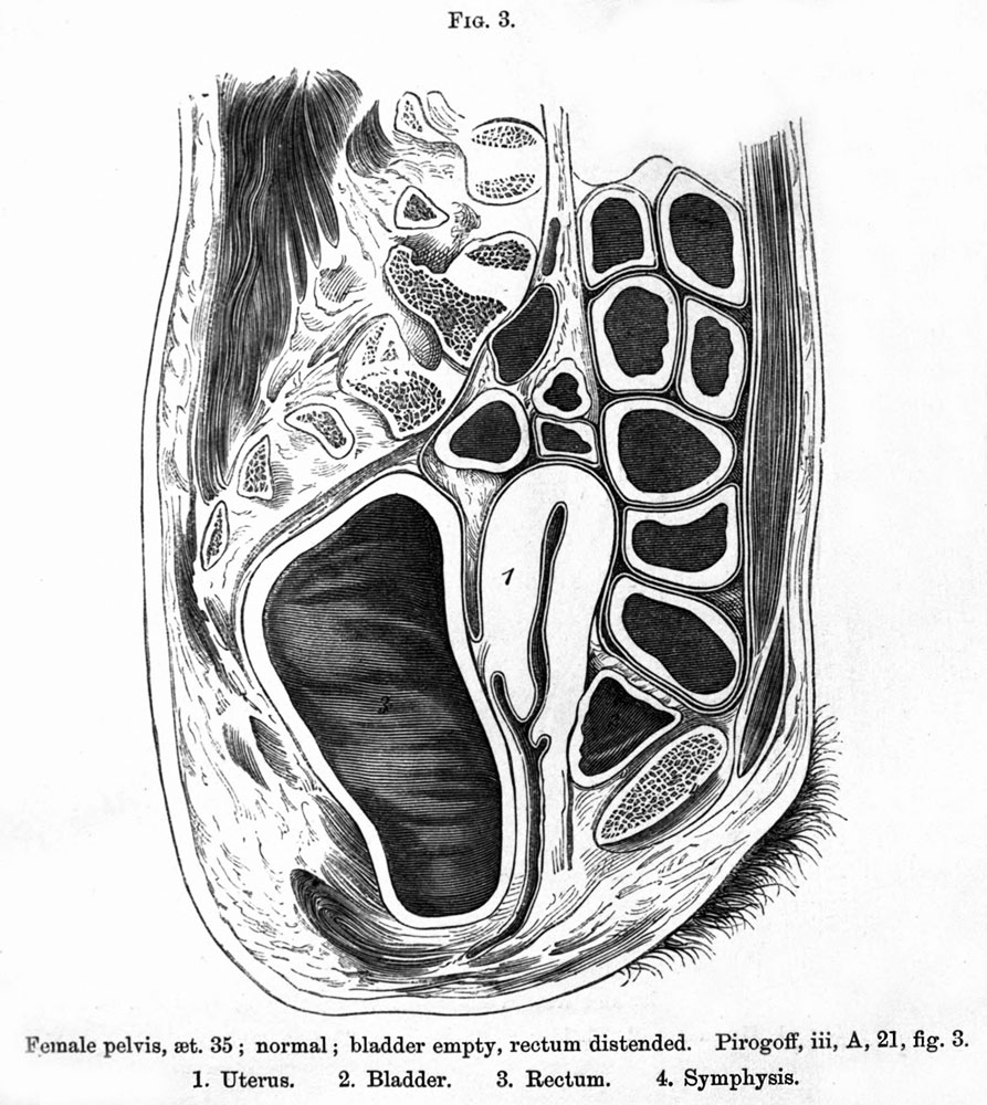 | 893 × 1,000 (208 KB) | Z8600021 (talk | contribs) | {{Braune 1877 header}} Braune 1877 plate 2 fig3.jpg |
You cannot overwrite this file.
File usage
The following 2 pages use this file: