File:Brain week 17 histology.jpg

Original file (1,050 × 697 pixels, file size: 61 KB, MIME type: image/jpeg)
Fetal Brain Development
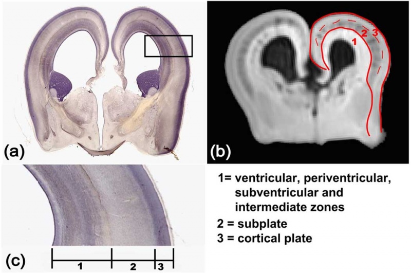
Layers of the developing cerebral cortex in the second trimester (week 17).
a–c - show a coronal histological slide of a 17 week fetal brain, a coronal MRI aDWI slice of a 17 week fetal brain, and an enlarged piece of the cerebral wall, respectively.
b - aDWI contrast clearly differentiates three layers that correspond to those regions in a and c.
The red contour is the boundary of the cortical plate and subplate (CP+SP). The dashed red curve separates the cortical plate and subplate.
The annotation of each layer is shown at the bottom right panel.
Original File Name: Figure 1 Nihms128503f1.jpg
Reference
<pubmed>19339620</pubmed>| PMC2721010 | J Neurosci.
Copyright: Copyright of all material published in The Journal of Neuroscience remains with the authors. The authors grant the Society for Neuroscience an exclusive license to publish their work for the first 6 months. After 6 months the work becomes available to the public to copy, distribute, or display under a Creative Commons Attribution-Noncommercial-Share Alike 3.0 Unported license.
File history
Click on a date/time to view the file as it appeared at that time.
| Date/Time | Thumbnail | Dimensions | User | Comment | |
|---|---|---|---|---|---|
| current | 11:33, 27 August 2010 |  | 1,050 × 697 (61 KB) | S8600021 (talk | contribs) | Layers of the developing cerebral cortex. a–c show a coronal histological slide of a 17 week fetal brain, a coronal MRI aDWI slice of a 17 week fetal brain, and an enlarged piece of the cerebral wall, respectively. In b, aDWI contrast clearly differ |
You cannot overwrite this file.
File usage
The following 14 pages use this file:
- 2011 Lab 12 - Second Trimester
- ANAT2341 Lab 11 - Second Trimester
- ANAT2341 Lab 12 - Second Trimester
- BGDA Practical 12 - Second Trimester
- Fetal Development
- Second Trimester
- Timeline human development
- Talk:Timeline human development
- Template:Second Trimester Timeline
- Template:Second Trimester Timeline collapsable table
- Template:Second Trimester table01
- Template talk:Second Trimester Timeline
- Category:Fetal
- Category:Second Trimester