File:Brain tract development 02.jpg: Difference between revisions
No edit summary |
|||
| Line 9: | Line 9: | ||
Original File Name: Figure 3 - 3D depiction of developmental white matter fibers. | Original File Name: Figure 3 - 3D depiction of developmental white matter fibers. | ||
{{Template:DTI}} | |||
==Reference== | ==Reference== | ||
Latest revision as of 15:15, 27 August 2010
Brain Tract Development - Limbic Tracts
3D depiction of developmental white matter fibers
A lateral view of limbic tracts where pink fibers in 13, 15, and 19 week brains are the fornix and stria terminalis and purple fibers in the 19 week brains indicate the cingulum bundle.
For anatomical guidance, the thalamus (yellow structure) and the ventricles (gray structure) are also shown.
Original File Name: Figure 3 - 3D depiction of developmental white matter fibers.
Diffusion tensor imaging (DTI) A newly developed form of magnetic resonance imaging (MRI). Magnetic field variations of the MRI magnet are applied in at least six different directions generating a three dimensional shape of the diffusion pattern. This technique can be used in neural imaging of white matter due to the orientation of axon bundles and the associated water flow. (More? Magnetic Resonance Imaging)
- Neural DTI Links: Scaled Fissures 13-21 weeks | Fissures 13-21 weeks | Brain Sylvian Fissure | Scaled Brain and Ventricles 13-21 weeks | Scaled Brain, Ventricles and Ganglia 13-21 weeks | Limbic Tract 13-19 weeks | Brain and Ventricles 13-21 weeks | Sylvian Fissure Movie | Neural System Development | Magnetic Resonance Imaging
Reference
<pubmed>19339620</pubmed>| PMC2721010 | J Neurosci.
Copyright: Copyright of all material published in The Journal of Neuroscience remains with the authors. The authors grant the Society for Neuroscience an exclusive license to publish their work for the first 6 months. After 6 months the work becomes available to the public to copy, distribute, or display under a Creative Commons Attribution-Noncommercial-Share Alike 3.0 Unported license.
File history
Click on a date/time to view the file as it appeared at that time.
| Date/Time | Thumbnail | Dimensions | User | Comment | |
|---|---|---|---|---|---|
| current | 11:57, 27 August 2010 | 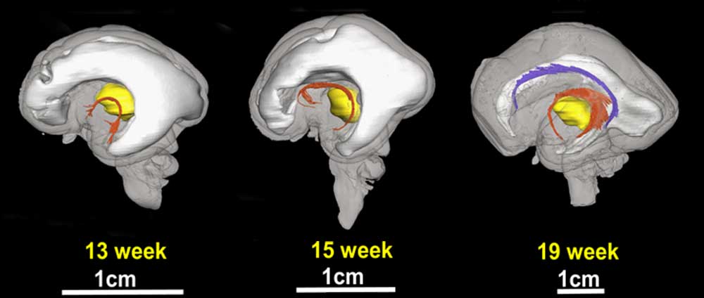 | 1,000 × 424 (29 KB) | S8600021 (talk | contribs) | ==Brain Tract Development - Limbic Tracts== 3D depiction of developmental white matter fibers '''a''' is a lateral view of limbic tracts where pink fibers in 13, 15, and 19 week brains are the fornix and stria terminalis and purple fibers in the 19 week |
You cannot overwrite this file.
File usage
The following page uses this file: