File:Brain Development.png: Difference between revisions
From Embryology
(Add reference and description) |
No edit summary |
||
| Line 1: | Line 1: | ||
==Description== | |||
==Reference== | |||
==Copyright== | |||
{{Template:Student Image}} | |||
Revision as of 12:17, 25 October 2017
Description
Reference
Copyright
- Note - This image was originally uploaded as part of an undergraduate science student project and may contain inaccuracies in either description or acknowledgements. Students have been advised in writing concerning the reuse of content and may accidentally have misunderstood the original terms of use. If image reuse on this non-commercial educational site infringes your existing copyright, please contact the site editor for immediate removal.
File history
Click on a date/time to view the file as it appeared at that time.
| Date/Time | Thumbnail | Dimensions | User | Comment | |
|---|---|---|---|---|---|
| current | 16:05, 25 October 2017 |  | 1,390 × 842 (1.92 MB) | Z5059949 (talk | contribs) | ==Description== Lateral perspective of the neural tube, showing the three flexures, and primary brain vesicles divided into the five subdivisions: Prosencephalon (Telencephalon and Diencephalon), Mesencephalon, and Rhombencephalon (Metencephalon, Myel... |
| 12:05, 25 October 2017 | 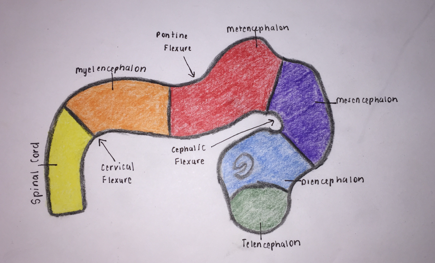 | 1,390 × 842 (1.92 MB) | Z5059949 (talk | contribs) | Add reference and description |
You cannot overwrite this file.
File usage
The following 2 pages use this file: