File:Brain-tract-development-04.jpg

Original file (1,000 × 424 pixels, file size: 30 KB, MIME type: image/jpeg)
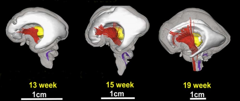
Brain Tract Development - projection fibers
3D depiction of developmental white matter fibers
A lateral view of projection fibers, where red and purple fibers in 13, 15, and 19 week brains are the cerebral peduncle and the inferior cerebellar peduncle, respectively.
d is a lateral view of association tracts, in which blue fibers in brains of 13 and 15 weeks are the external capsule, and green and red fibers in a brain of 19 weeks are the inferior longitudinal fasciculus/inferior fronto-occipital peduncle and uncinate fasciculus, respectively.
For anatomical guidance, the thalamus (yellow structure) and the ventricles (gray structure) are also shown.
Original File Name: Figure 3 - 3D depiction of developmental white matter fibers.
Reference
<pubmed>19339620</pubmed>| PMC2721010 | J Neurosci.
Copyright: Copyright of all material published in The Journal of Neuroscience remains with the authors. The authors grant the Society for Neuroscience an exclusive license to publish their work for the first 6 months. After 6 months the work becomes available to the public to copy, distribute, or display under a Creative Commons Attribution-Noncommercial-Share Alike 3.0 Unported license.
File history
Click on a date/time to view the file as it appeared at that time.
| Date/Time | Thumbnail | Dimensions | User | Comment | |
|---|---|---|---|---|---|
| current | 12:13, 27 August 2010 | | 1,000 × 424 (30 KB) | S8600021 (talk | contribs) | ==Brain Tract Development - projection fibers== 3D depiction of developmental white matter fibers A lateral view of projection fibers, where red and purple fibers in 13, 15, and 19 week brains are the cerebral peduncle and the inferior cerebellar pedunc |
You cannot overwrite this file.
File usage
The following page uses this file: