File:Bradley1908 fig02.jpg: Difference between revisions
mNo edit summary |
(Z8600021 uploaded a new version of File:Bradley1908 fig02.jpg) |
(No difference)
| |
Latest revision as of 21:02, 21 November 2019
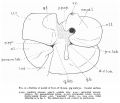
Fig. 2. Outline of model of liver of 15-mm. pig embryo
Caudal surface.
p.pap., papillary process; caud.l., caudate lobe; p.pyr., pyramidal process ; paraum. lob., paraumbilical lobule; p-c.lob., precaudate lobule; d-v.lob., dextro-vesical lobule; y.lob., ‘‘quadrate” lobule; v.c., vena cava. Other lettering as in fig. 1. The dotted area is not covered by peritoneum.
Reference
Bradley OC. A contribution to the morphology and development of the mammalian liver. (1908) J Anat. 43: 1-42. PMID 17232788
Cite this page: Hill, M.A. (2024, April 19) Embryology Bradley1908 fig02.jpg. Retrieved from https://embryology.med.unsw.edu.au/embryology/index.php/File:Bradley1908_fig02.jpg
- © Dr Mark Hill 2024, UNSW Embryology ISBN: 978 0 7334 2609 4 - UNSW CRICOS Provider Code No. 00098G
File history
Click on a date/time to view the file as it appeared at that time.
| Date/Time | Thumbnail | Dimensions | User | Comment | |
|---|---|---|---|---|---|
| current | 21:02, 21 November 2019 |  | 1,000 × 714 (75 KB) | Z8600021 (talk | contribs) | |
| 20:59, 21 November 2019 |  | 1,000 × 859 (99 KB) | Z8600021 (talk | contribs) |
You cannot overwrite this file.
File usage
The following 2 pages use this file: