File:Boyd1950 fig06.jpg: Difference between revisions
(Z8600021 uploaded a new version of File:Boyd1950 fig06.jpg) |
|||
| Line 1: | Line 1: | ||
==Fig. 6. Sections through developing fourth pouch of human embryos== | ==Fig. 6. Sections through developing fourth pouch of human embryos== | ||
To show relationship between | To show relationship between “ventral component ” of this pouch and the differentiating thyroid tissue. (x 450.) | ||
(a) Left side 13 mm. human embryo | |||
(b) Right side 17 mm. human embryo; | |||
(c) Left side 17 mm. human embryo | |||
(a') Left side 19 mm. human embryo. | |||
Note different appearance presented by the “ventral component” of the fourth pouch. In (b) and (c) it appears to be more intimately related to median {{thyroid}} tissue than in the other sections. | |||
{{Boyd1950 figures}} | {{Boyd1950 figures}} | ||
| Line 9: | Line 19: | ||
{{Footer}} | {{Footer}} | ||
[[Category:Thymus]][[Category:Fetal]][[Category:Parathyroid]] | [[Category:Thymus]][[Category:Fetal]][[Category:Parathyroid]][[Category:Thyroid]] | ||
Latest revision as of 11:30, 30 July 2020
Fig. 6. Sections through developing fourth pouch of human embryos
To show relationship between “ventral component ” of this pouch and the differentiating thyroid tissue. (x 450.)
(a) Left side 13 mm. human embryo
(b) Right side 17 mm. human embryo;
(c) Left side 17 mm. human embryo
(a') Left side 19 mm. human embryo.
Note different appearance presented by the “ventral component” of the fourth pouch. In (b) and (c) it appears to be more intimately related to median thyroid tissue than in the other sections.
| Historic Disclaimer - information about historic embryology pages |
|---|
 |
- Links: fig 1 | fig 2 | fig 3a | fig 3b | fig 4 | fig 5 | fig 6 | fig 7 | fig 8 | fig 9 | fig 10 | 1950 Boyd | Thyroid | Parathyroid | Thymus | Historic Papers
Reference
Boyd JD. Development of the thyroid and parathyroid glands and the thymus. (1950) Ann R Coll Surg Engl. 7(6): 455-71. PMID 14790564
Cite this page: Hill, M.A. (2024, April 19) Embryology Boyd1950 fig06.jpg. Retrieved from https://embryology.med.unsw.edu.au/embryology/index.php/File:Boyd1950_fig06.jpg
- © Dr Mark Hill 2024, UNSW Embryology ISBN: 978 0 7334 2609 4 - UNSW CRICOS Provider Code No. 00098G
File history
Click on a date/time to view the file as it appeared at that time.
| Date/Time | Thumbnail | Dimensions | User | Comment | |
|---|---|---|---|---|---|
| current | 07:37, 21 March 2017 | 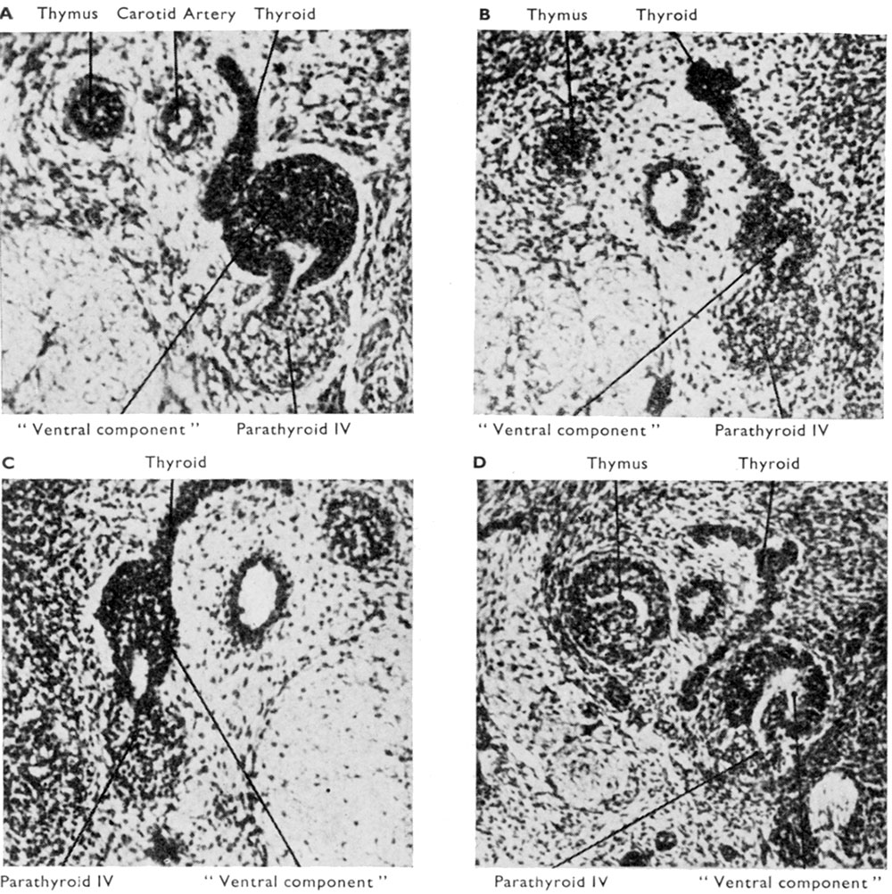 | 1,000 × 1,002 (272 KB) | Z8600021 (talk | contribs) | |
| 07:36, 21 March 2017 |  | 1,347 × 1,606 (666 KB) | Z8600021 (talk | contribs) |
You cannot overwrite this file.
File usage
The following page uses this file: