File:Boyd1950 fig03b.jpg
From Embryology

Size of this preview: 361 × 599 pixels. Other resolution: 600 × 996 pixels.
Original file (600 × 996 pixels, file size: 152 KB, MIME type: image/jpeg)
Fig. 3B. Coronal section through parathyroid III and thymus of a 48 mm human embryo
The main bulk of the thymus has now separated from parathyroid II], but some thymic tissue is shown still connected with the parathyroid. On the right side of - this aberrant thymic tissue a Kürsteiner canal is shown.
File history
Click on a date/time to view the file as it appeared at that time.
| Date/Time | Thumbnail | Dimensions | User | Comment | |
|---|---|---|---|---|---|
| current | 22:43, 6 March 2017 | | 600 × 996 (152 KB) | Z8600021 (talk | contribs) | |
| 22:43, 6 March 2017 | 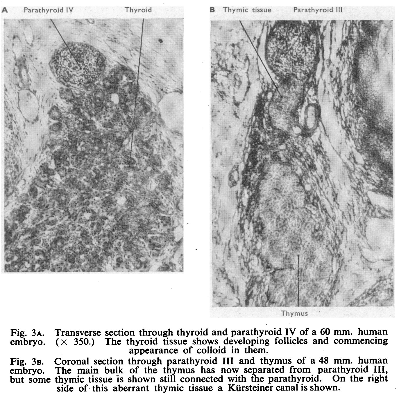 | 1,373 × 1,359 (566 KB) | Z8600021 (talk | contribs) |
You cannot overwrite this file.
File usage
The following page uses this file: