File:Boyd1950 fig03b.jpg: Difference between revisions
From Embryology
mNo edit summary |
mNo edit summary |
||
| Line 7: | Line 7: | ||
{{Footer}} | {{Footer}} | ||
[[Category:Thymus]][[Category:Fetal]] | |||
Revision as of 07:14, 21 March 2017
Fig. 3B. Coronal section through parathyroid III and thymus of a 48 mm human embryo
The main bulk of the thymus has now separated from parathyroid II], but some thymic tissue is shown still connected with the parathyroid. On the right side of - this aberrant thymic tissue a Kürsteiner canal is shown.
Reference
Boyd JD. Development of the thyroid and parathyroid glands and the thymus. (1950) Ann R Coll Surg Engl. 7(6): 455-71. PMID 14790564
Cite this page: Hill, M.A. (2024, April 20) Embryology Boyd1950 fig03b.jpg. Retrieved from https://embryology.med.unsw.edu.au/embryology/index.php/File:Boyd1950_fig03b.jpg
- © Dr Mark Hill 2024, UNSW Embryology ISBN: 978 0 7334 2609 4 - UNSW CRICOS Provider Code No. 00098G
File history
Click on a date/time to view the file as it appeared at that time.
| Date/Time | Thumbnail | Dimensions | User | Comment | |
|---|---|---|---|---|---|
| current | 22:43, 6 March 2017 |  | 600 × 996 (152 KB) | Z8600021 (talk | contribs) | |
| 22:43, 6 March 2017 | 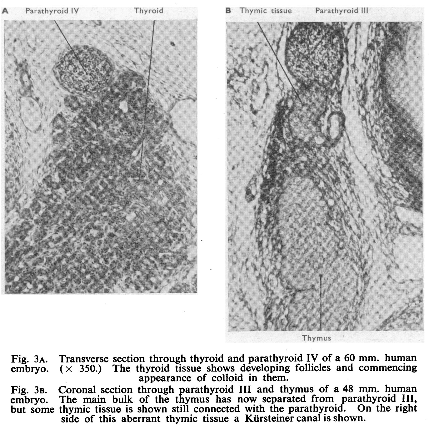 | 1,373 × 1,359 (566 KB) | Z8600021 (talk | contribs) |
You cannot overwrite this file.
File usage
The following page uses this file: