File:Bonnot1906 fig03.jpg

Original file (1,192 × 1,689 pixels, file size: 154 KB, MIME type: image/jpeg)
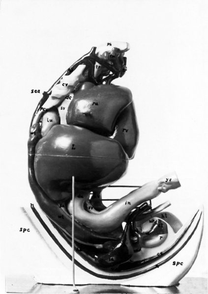
Fig. 3. From a photograph of the right view of the model
On this side, the lower part of the body wall is not seen as on the left side, but has been dissected out to the mid-sagittal plane, giving a side view of the spinal cord, notochord, the descending aorta with its branches, and the pelvic viscera.
Explanation
Explanation (in addition to letters shown in Fig. 2): a‘ right aortic arch. al allantois. b Wolffian duet. ca caudal artery. cl cloaca. d duodenum. ha hypogastrie artery. i intestine. n notochord. o gall bladder. r rectum. ra right auricle. rv right ventricle. sc spinal cord. sv sinus venosus. u ureter. Its T-shaped upper extremity, forming the anlage of the permanent kidney, is almost entirely hidden by the hypogastric artery. up urinogenital papilla. vv vitelline (omphalomesenteric) vein.
|
A ascending aorta. a dorsal aorta. a‘ left aortic arch. ac anterior cardinal vein (sinus-like dilatation). c caecum, not marked externally, though its cavity is distinct internally. co colon. d ductus Cuvier. hl hind limb. l lung. L liver. la left auricle. lv left ventricle. |
m mesentery. pa posterior cardinal vein. ph pharynx. s somite (external surface). so anlage of sexual gland. sc origin of subclavian artery. sr suprarenal body (slightly visible). ta tail. th thymus, including the main gland and also the smaller “nodulus thymicus”. tl lateral anlage of the thyroid gland, located between the fourth and fifth branehial arterial arches. tm, median thyroid anlage. |
u umbilical vein. w Wolffian body. x quadrangular window cut through the thick-walled great omentum into the bursa omentalis. The anlage of the imperfectly differentiated spleen lies in the oiuental wall just behind this window. Internal to the omentum lies the stomach (visible through the window in the original model, though not in the photograph). ys yolk stalk, out near attachment to intestinal loop. |
| Week: | 1 | 2 | 3 | 4 | 5 | 6 | 7 | 8 |
| Carnegie stage: | 1 2 3 4 | 5 6 | 7 8 9 | 10 11 12 13 | 14 15 | 16 17 | 18 19 | 20 21 22 23 |
Reference
Bonnet E. and Severs R. On the structure of a human embryo eleven millimeters in length. (1906) Anat. Anz., 29: 452-459.
Cite this page: Hill, M.A. (2024, April 23) Embryology Bonnot1906 fig03.jpg. Retrieved from https://embryology.med.unsw.edu.au/embryology/index.php/File:Bonnot1906_fig03.jpg
- © Dr Mark Hill 2024, UNSW Embryology ISBN: 978 0 7334 2609 4 - UNSW CRICOS Provider Code No. 00098G
File history
Click on a date/time to view the file as it appeared at that time.
| Date/Time | Thumbnail | Dimensions | User | Comment | |
|---|---|---|---|---|---|
| current | 16:44, 19 December 2016 | | 1,192 × 1,689 (154 KB) | Z8600021 (talk | contribs) | ===Reference=== {{Ref-Bonnot1906}} {{Footer}} Category:Carnegie Stage 16Category:Week 6 Category:1900's |
You cannot overwrite this file.
File usage
The following 2 pages use this file: