File:Bladder histology 01.jpg
Bladder_histology_01.jpg (480 × 600 pixels, file size: 29 KB, MIME type: image/jpeg)
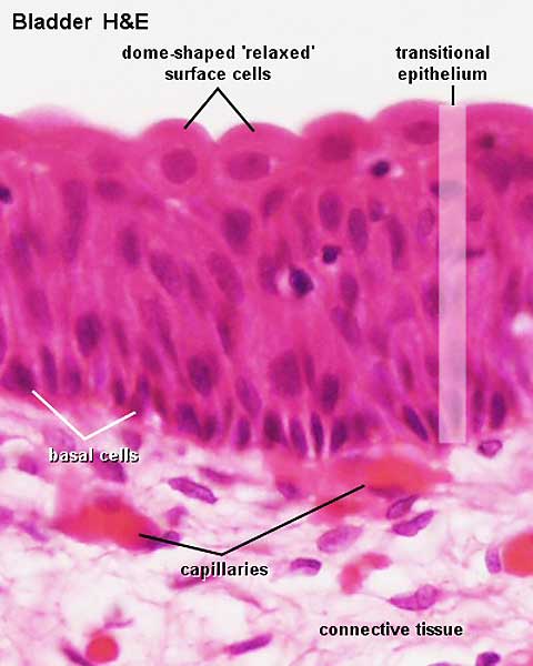
Bladder Histology
(Stain - Haematoxylin Eosin)
Transitional epithelium is found exclusively in the excretory urinary passages.
- renal calyces and pelvis
- ureter
- urinary bladder
- part of the urethra
The shape of the surface cells and the number of rows change if the bladder is distended (number of rows decreases).
- Renal Histology: Histology | Histology Stains | Renal Development
- Kidney - Nephron overview | Glomerulus | Vascular and renal poles | Medullary ray | tubules
- Ureter - Ureter labeled | Ureter epithelium
- Bladder - overview | wall 1 | wall 2 | transitional epithelium | Urinary Bladder Development
Links: Histology | Histology Stains | Blue Histology images copyright Lutz Slomianka 1998-2009. The literary and artistic works on the original Blue Histology website may be reproduced, adapted, published and distributed for non-commercial purposes. See also the page Histology Stains.
Cite this page: Hill, M.A. (2024, April 25) Embryology Bladder histology 01.jpg. Retrieved from https://embryology.med.unsw.edu.au/embryology/index.php/File:Bladder_histology_01.jpg
- © Dr Mark Hill 2024, UNSW Embryology ISBN: 978 0 7334 2609 4 - UNSW CRICOS Provider Code No. 00098G
File history
Click on a date/time to view the file as it appeared at that time.
| Date/Time | Thumbnail | Dimensions | User | Comment | |
|---|---|---|---|---|---|
| current | 14:31, 25 March 2012 | | 480 × 600 (29 KB) | Z8600021 (talk | contribs) | ==Bladder Histology== Transitional epithelium is found exclusively in the excretory urinary passages. * renal calyces and pelvis * ureter * urinary bladder * part of the urethra {{Blue Histology}} |
You cannot overwrite this file.
File usage
The following 3 pages use this file: