File:BaxterBoyd1939-text-fig02.jpg: Difference between revisions
(Z8600021 uploaded a new version of File:BaxterBoyd1939-text-fig02.jpg) |
|||
| Line 1: | Line 1: | ||
==Text- | ==Text-Fig. 2. Diagram to show the arrangement of the acoustico-facial neural crest primordia in certain human embryos== | ||
Ectoderm, neural folds and tube indicated in solid black; acoustico-facial neural crest material stippled; endoderm and dorsal aortae outlined. (For explanation see text below) | |||
* '''A.''' Payne embryo of seven somites. | * '''A.''' Payne embryo of seven somites. | ||
| Line 7: | Line 8: | ||
The form taken by the acoustico-facial neural crest mass in this embryo is very different from that seen in Corner’s ten-somite human embryo (cp. Text-fig. 2B). In his specimen the anterior limit of fusion of the neural folds has not extended so far forward as in our embryo. It reaches only to the level of the acoustico-facial neural crest primordium which, here, is represented by bilateral and entirely separate cell aggregations; these, according to Corner’s description and microphotographs, seem to stream out of the angle of junction of the neural and somatic ectoderm. In some regions he found a sharp demarcation of the neural crest cells from the adjacent mesenchyme; in other regions it was impossible to draw a line between the lateral margin of the crest and the mesodermal elements. In our embryo we find no difficulty in distinguishing between the cells of the acoustico-facial primordium and the surrounding, frankly mesodermal, elements. Hence there is no appearance of continuity between the crest material and the hyoid, or other, branchial mesodermal condensations. Other human embryos of the somite period in which the neural crests have been studied seem to correspond with Corner’s description of the arrangement of the acoustico-facial neural crest elements. | |||
Thus in Payne’s (1925) embryo of seven somites (Text-fig. 2A), and Veit’s embryo of eight somites, there are two, quite separate, acoustico-facial primordia; and Heuser (1930), in his fourteen-somite specimen, describes bilateral acoustico-facial primordia in an embryo with complete closure of the neural tube in the definitive hindbrain region (Text-fig. 2C). | |||
{{BaxterBoyd1939 figures}} | {{BaxterBoyd1939 figures}} | ||
Revision as of 11:57, 19 May 2017
Text-Fig. 2. Diagram to show the arrangement of the acoustico-facial neural crest primordia in certain human embryos
Ectoderm, neural folds and tube indicated in solid black; acoustico-facial neural crest material stippled; endoderm and dorsal aortae outlined. (For explanation see text below)
- A. Payne embryo of seven somites.
- B. Corner embryo (ten somites).
- C. Heuser embryo (fourteen somites).
- D. Baxter-Boyd embryo (ten somites).
The form taken by the acoustico-facial neural crest mass in this embryo is very different from that seen in Corner’s ten-somite human embryo (cp. Text-fig. 2B). In his specimen the anterior limit of fusion of the neural folds has not extended so far forward as in our embryo. It reaches only to the level of the acoustico-facial neural crest primordium which, here, is represented by bilateral and entirely separate cell aggregations; these, according to Corner’s description and microphotographs, seem to stream out of the angle of junction of the neural and somatic ectoderm. In some regions he found a sharp demarcation of the neural crest cells from the adjacent mesenchyme; in other regions it was impossible to draw a line between the lateral margin of the crest and the mesodermal elements. In our embryo we find no difficulty in distinguishing between the cells of the acoustico-facial primordium and the surrounding, frankly mesodermal, elements. Hence there is no appearance of continuity between the crest material and the hyoid, or other, branchial mesodermal condensations. Other human embryos of the somite period in which the neural crests have been studied seem to correspond with Corner’s description of the arrangement of the acoustico-facial neural crest elements.
Thus in Payne’s (1925) embryo of seven somites (Text-fig. 2A), and Veit’s embryo of eight somites, there are two, quite separate, acoustico-facial primordia; and Heuser (1930), in his fourteen-somite specimen, describes bilateral acoustico-facial primordia in an embryo with complete closure of the neural tube in the definitive hindbrain region (Text-fig. 2C).
| Historic Disclaimer - information about historic embryology pages |
|---|
 |
- Links: Text-fig 1 | Text-fig 2 | Plate 1 | Fig 1 | Fig 2 | Fig 3 | Fig 4 | Plate 2 | Fig 5 | Fig 6 | Fig 7 | Baxter and Boyd 1939 | Historic Embryology Papers | Neural Crest Development | Carnegie stage 10
Reference
Baxter JS. and Boyd JD. Observations on the neural crest of a ten-somite human embryo. (1939) J Anat. 73: 318–326. PMID 17104759
Cite this page: Hill, M.A. (2024, April 19) Embryology BaxterBoyd1939-text-fig02.jpg. Retrieved from https://embryology.med.unsw.edu.au/embryology/index.php/File:BaxterBoyd1939-text-fig02.jpg
- © Dr Mark Hill 2024, UNSW Embryology ISBN: 978 0 7334 2609 4 - UNSW CRICOS Provider Code No. 00098G
File history
Click on a date/time to view the file as it appeared at that time.
| Date/Time | Thumbnail | Dimensions | User | Comment | |
|---|---|---|---|---|---|
| current | 18:02, 16 September 2015 | 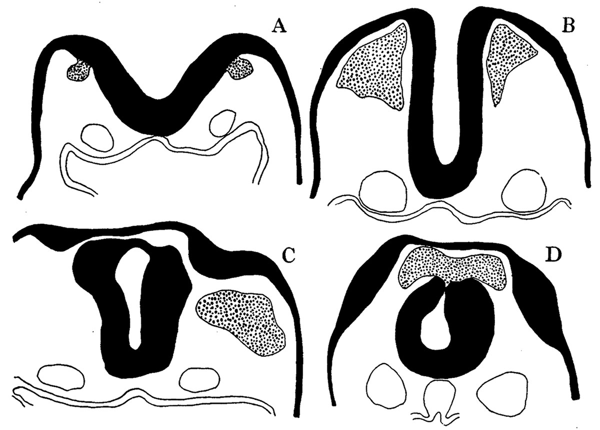 | 1,200 × 861 (133 KB) | Z8600021 (talk | contribs) | |
| 18:00, 16 September 2015 |  | 1,438 × 1,234 (298 KB) | Z8600021 (talk | contribs) |
You cannot overwrite this file.
File usage
The following 2 pages use this file: